

These images are provided to foster education about Johne’s disease. They are distributed under the terms of the Creative Commons Attribution License which permits unrestricted use, distribution, and reproduction in any medium, provided the source, johnes.org, is credited.
If higher resolution versions of these images are required, please contact the site author: M.T. Collins. Commercial users may be asked to pay a fee for commercial use of high quality images.
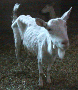 Goats with clinical Johne’s disease most often looks like a poorly fed, unthrifty goat. Animals that produce fecal pellets, such as goats, generally do not develop diarrhea, although the feces may become pasty like dog feces. Although appearing sickly, goats with Johne’s disease continue eating well. The goat pictured herd is a Saanen, a popular breed of goats used for milk production. Sorry about the poor lighting, the photo was taken in the barn. Photo taken on a dairy farm in southern Chile by M.T. Collins while on sabbatical at Universidad Austral de Chile.
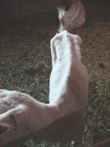 This is the top view of the goat pictured above to better illustrate the poor body condition of the animal. This herd of goats was intended to be a source of genetics for local dairy goat herds in hopes of increasing milk production. Sadly, these goats would bring MAP into other goats herds along with the better genetics. This is a recurring theme in how Johne’s disease spreads and emphasizes why breeders who sell animals of high genetic merit must be free of MAP.
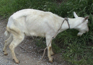 This better image of the Chilean goat with Johne’s disease is shown here. More about the herd and the accuracy of ELISAs in goats can be found in the publication by Salgado et al. Journal of Veterinary Diagnostic Investigation 19:99-102, 2007.
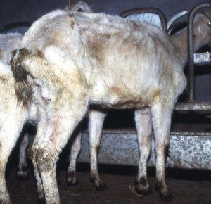
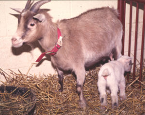 This pygmy goat was a member of a herd of 30 goats in Minnesota with a high rate of Johne’s disease. Several of the goats were donated to the University of Wisconsin to aid in the study of this disease in small goat breeds. The goat pictured here was in the advanced stages of a MAP infection and thin, although this was not apparent without feeling the goat and noting the prominent ribs. The goat had a kid and within one month died from it’s MAP infection.
In the months before death on August 3, 1995, this pygmy goat had a steadily rising level of serum antibody and was consistently fecal culture-positive (PCR was not yet available).
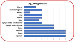 A necropsy was performed and multiple tissues were quantitatively cultured for MAP. MAP was recovered from 12 tissues in numbers ranging from 10 to 10,000,000 MAP per gram of tissue as shown on the adjacent graphic. These data illustrate how MAP infections eventually become disseminated throughout the body, i.e. a systemic, or generalized infection. This also explains why MAP is found in milk and meat from animals with a last-stage MAP infection.
Read more about this outbreak in the 2003 publication by Manning et al.
 Dairy goat with clinical Johne’s disease. Photo by M.T. Collins taken in a herd in The Netherlands.