

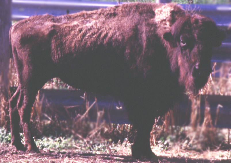
American Bison (Bison bison) are susceptible to MAP. Outbreaks have been documented in multiple herds in the U.S. and Canada. The clinical presentation is just low body condition with diarrhea being uncommon.
Most of our knowledge regarding Johne’s disease in Bison come from a study of one outbreak as reported by Buergelt et al. (The pathology of spontaneous paratuberculosis in North American Bison (Bison bison), Veterinary Pathology 37:428-438, 2000). The following are excerpts from that Open Access publication:
Gross and histopathologic examinations were performed on 70 North American bison (Bison bison) from a Mycobacterium avium paratuberculosis culture–positive herd. The bison examined were part of a breeding herd totaling 2,800 animals. Of the examined bison, the gross changes consistent with paratuberculosis included weight loss due to skeletal muscle mass reduction, atrophy of body fat, or both, in 28 of 70 (40%) animals; thickening of the mucosa of the small intestine in 8 of 70 (11%) animals; and prominent mesenteric or ileocecal lymph nodes in 16 of 70 (23%) animals. One animal had distended serosal lymphatics. Histologic lesions compatible with Johne’s disease were diagnosed in 30 of 70 (43%) bison on the basis of the demonstration of noncaseating granulomatous inflammatory infiltrates and of one or more acid-fast bacilli characteristic of Mycobacterium avium paratuberculosis. A suspicious diagnosis of Johne’s disease was obtained in 11 of 70 (16%) bison on the basis of the observation of noncaseating granulomatous inflammatory infiltrates without demonstrable acid-fast bacteria. Twenty-nine of 70 (41%) animals were assessed as histologically paratuberculosis free.
Multiple diagnostic tests were used on these same Bison with these findings: The AGID test identified only one (3%) of the 30 mophologically positive bison, whereas the ELISA test, after some modification, identified 19 (63%) of the morphologically positive bison. Only 25 bison were subjected to fecal culture, and 8 of the 20 (40%) of the morphologically positive bison were correctly identified. One bison morphologically classified as suspicious had a positive fecal culture. The same 25 bison were also subjected to tissue culture, and 12 of 20 (60%) of them were correctly identified. Finally, 49 bison were subjected to a PCR test on feces, and 50 were subjected to a tissue PCR test. The fecal PCR identified 22 of 30 (73%) of the morphologically positive bison correctly, but it also identified eight additional bison as positive when the other tests, including the histologic tests, were negative in these animals. The tissue PCR identified 100% of the positive bison, but it also identified 15 of the suspicious and negative bison as positive.
From the comparison of these results, we concluded that the incidence of paratuberculosis infection in bison was higher when using PCR testing than histopathology or other tests. Some of the tests correlated with each other, whereas others did not. In some circumstances, acid-fast bacteria were microscopically detected in the tissue of bison, but the organisms could not be cultured from feces or tissues, and specific antibodies could not be detected from serum samples. This suggests that tests that work for cattle need to be modified to make them work reliably for bison. The discrepancy between tests prompted the overall conclusion that the results of attempts to diagnose paratuberculosis in bison with the test methods usually applied to cattle do not follow the pattern of sensitivity and specificity expected from these tests in cattle.