

These images are provided to foster education about Johne’s disease. They are distributed under the terms of the Creative Commons Attribution License which permits unrestricted use, distribution, and reproduction in any medium, provided the source, johnes.org, is credited.
If higher resolution versions of these images are required, please contact the site author: M.T. Collins. Commercial users may be asked to pay a fee for commercial use of high quality images.
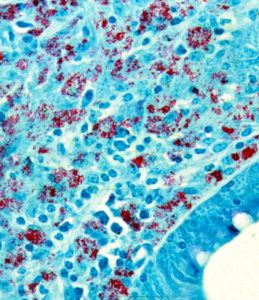 Bovine ileum tissue section with Ziehl-Neelson acid-fast stain magnified ~400 times. MAP bacteria (red) are clustered inside macrophages. Mononuclear inflammatory cells, macrophages and lymphocytes, surround the infected macrophages. In cattle with late-stage Johne’s disease, MAP bacteria are typically numerous as shown here. This is the so called “multibacillary” or “lepromatous” (meaning similar to advanced forms of leprosy) type of infection. The host response is called diffuse granulomatous inflammation, similar to what is seen in human leprosy and Crohn’s disease (although acid-fast bacteria are generally not seen in Crohn’s disease). This inflammation is different from what is seen in tuberculosis where the inflammation is organized into a walled-off granuloma.
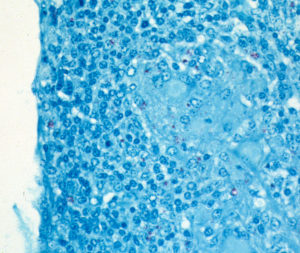 Bovine mesenteric lymph node section stained with Ziehl-Neelsen acid-fast stain (magnified 250x) showing the tiny red-colored MAP bacteria inside macrophages. Look closely and see below for a higher magnification.
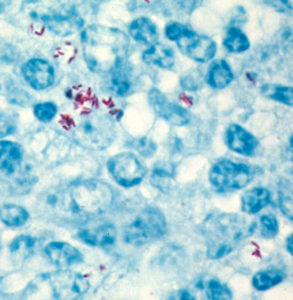 High power (1,000x) magnification of the image above showing how important it is to use special, acid-fast, stains and to look at the histopathology at magnification provided by an oil immersion lens to clearly see MAP bacteria in lesions.
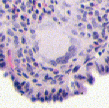
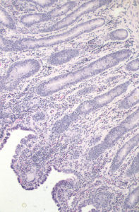 H&E stained section of bovine intestine showing the granulomatous inflammation characteristic of Johne’s disease.
 Multi-nucleated giant cell commonly found in the granulomatous inflammation caused by MAP in cattle.