

These images are provided to foster education about Johne’s disease. They are distributed under the terms of the Creative Commons Attribution License which permits unrestricted use, distribution, and reproduction in any medium, provided the source, johnes.org, is credited.
If higher resolution versions of these images are required, please contact the site author: M.T. Collins. Commercial users may be asked to pay a fee for commercial use of high quality images.
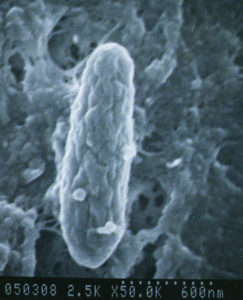
This is a portion of a larger scanning electron micrograph. It is one of several images created by Rebecca Van Boxtel working in the laboratory of Michael T. Collins. Her studies concerned the influence of polyoxyethylene sorbate compounds, commonly called Tween, on the morphology of MAP cells and MAP cultures. For more details, see R.M. Van Boxtel, R.S. Lambrecht, and M.T. Collins. 1990. APMIS 98:901-908,1990.
Without Tween, MAP has a rough cell surface. Tween causes cells to look smooth. Colonies of MAP growing on solid media are likewise rough or smooth depending on the presence of Tween.
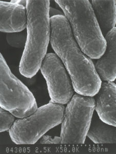
 Scanning electron micrograph of rough MAP grown in culture media without Tween. Perhaps the most popular image in our collection, it is one of several images created by Rebecca Van Boxtel working in the laboratory of Michael T. Collins. Her studies concerned the influence of polyoxyethylene sorbate compounds, commonly called Tween, on the morphology of MAP cells and MAP cultures. For more details, see R.M. Van Boxtel, R.S. Lambrecht, and M.T. Collins. 1990. APMIS 98:901-908,1990.
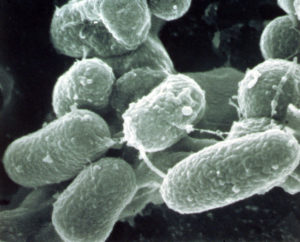
 When grown in the presence of Tween, MAP has a smooth cell surface. Colonies of MAP growing on solid media are likewise rough or smooth depending on the presence of Tween (see below). The relevance for researchers is that rough and smooth cells may behave differently when infecting macrophages in vitro or in animal challenge trials. This image was created by Rebecca Van Boxtel working in the laboratory of Michael T. Collins.
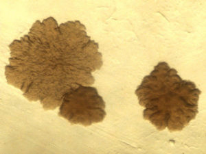 Rough-looking colonies of MAP bacteria originating from a cow when grown on Middlebrook 7H9 agar without Tween 80. Tween 80 significantly increases the rate of growth of MAP in broth media. This image was created by Rebecca Van Boxtel working in the laboratory of Michael T. Collins. For more details, see R.M. Van Boxtel, R.S. Lambrecht, and M.T. Collins. 1990. APMIS 98:901-908,1990.
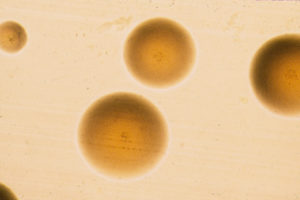 Smooth-looking colonies of MAP bacteria originating from a cow from when grown on Middlebrook 7H9 agar with 1.0% Tween 80. This image was created by Rebecca Van Boxtel working in the laboratory of Michael T. Collins.
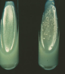 Herrold’s Egg Yolk (HEY) agar. The standard solid culture medium used to growth MAP from clinical samples such as feces. MAP will only grow on this culture medium only when it is supplemented with mycobactin-J (see tube on the right-hand side). Failure to grow MAP on this same culture medium without mycobactin-J (left-hand tube) is one of the unique characteristics of MAP that distinguishes it from most all other mycobacteria. Many laboratories still use HEY to diagnosis Johne’s disease by fecal culture. Liquid culture systems, BACTEC-MGIT and Trek-ESP, have replaced HEY in many because labs because MAP growth can be detected more quickly, i.e. in 4-8 weeks instead of 8-12 weeks on HEY.
 This composite image highlights the extreme difference in appearance of MAP grown in liquid 7H9 broth compared to Watson-Reid broth culture medium. The top half of image shows MAP in 7H9: visible growth floating on the broth medium surface the left and a scanning electron micrograph of the smoother-looking MAP bacteria on the right. The lower half of image shows MAP in Watson-Reid medium: visible wrinkled surface growth on the left and a scanning electron micrograph on the rougher looking MAP bacteria on the right. Culture medium composition has a profound effects on the chemistry and physiology of MAP. For more on this, see N. Sung and M.T. Collins, Applied and Environmental Microbiology 69:6833-6840, 2003.