

These images are provided to foster education about Johne’s disease. They are distributed under the terms of the Creative Commons Attribution License which permits unrestricted use, distribution, and reproduction in any medium, provided the source, johnes.org, is credited.
If higher resolution versions of these images are required, please contact the site author: M.T. Collins. Commercial users may be asked to pay a fee for commercial use of high quality images.
Bovine (Cattle) Gross Pathology
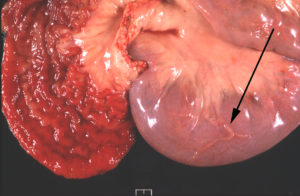 Classical lesions are a thickened, corrugated mucosal surface of the ileum and dilated lymphatics on the serosal surface (arrow on adjacent image). This exceptional photo was taken by Dr. A.J. Cooley and has appeared in several publications.
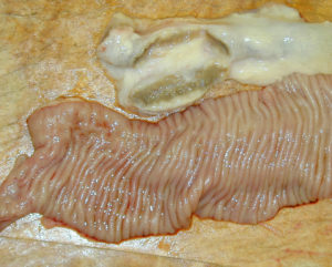
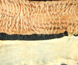 This photo provided by J.B. Jorgensen, State Veterinary Serum Laboratory, Copenhagen, Denmark, shows the thickened and corrugated mucosal surface of the ileum (top) in contrast with a normal ileum (bottom).
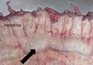
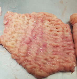 Photo taken by a veterinary student, named Stephanie, during a clinical rotation in a U.S. slaughterhouse. She sent me this photo and indicated that it was very common to see cattle with this obviously thickened intestine.
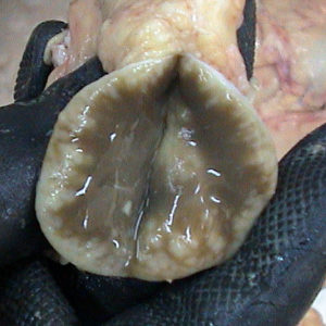
 Prominent Peyer’s Patches on the surface of the ileum (raised slightly red tissue in the center) from a cow wit clinical Johne’s disease.
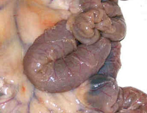
 Prominent mesenteric lymph nodes (arrow) from an Angus cow with clinical Johne’s disease.
Caprine (Goat) Gross Pathology
 Cross section of an enlarged lymph node near the intestinal tract. Such lymph nodes are typically smaller and on cross section a uniform brownish liver-color. This lymph node is enlarged and has accumulations of while blood cells around the periphery of the lymph node indicative of the inflammatory response taking place here.
In goats, thickening of the intestinal wall is not as pronounced as it is in cattle with Johne’s disease.
Ovine (Sheep) Gross Pathology
![]() Prominent mesenteric lymph nodes (arrow) adjacent to the small intestines of a sheep with clinical Johne’s disease (photo by Dr. Suelee Rob-Austerman).
Prominent mesenteric lymph nodes (arrow) adjacent to the small intestines of a sheep with clinical Johne’s disease (photo by Dr. Suelee Rob-Austerman).
 Serosal (external) surface of the small intestine of a sheep with clinical Johne’s disease showing the prominent lymphatic vessels (thin white lines).
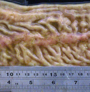
 Thickened and corrugated ileum of a sheep with clinical Johne’s disease. Intestinal thickening is less dramatic and less often seen in sheep as compared to cattle. This is an excellent image was provided by Dr. by Suelee Rob-Austerman.