

Johne’s disease has been diagnosed in multiple species of free-ranging, wild, ruminants. Although not definitively proven, the opinion of experts is that in most situations MAP infections in domestic livestock are spilling over into wildlife and that the resulting MAP infections in these wildlife compromise the health of these free-ranging animals but are not a significant risk to domestic livestock. A possible exception is the relationship of MAP-infected rabbits in Scotland (see non-ruminants) and the animals sharing pastures with them.
Below are listed just some of the more significant and well-documented occurrences of Johne’s disease in free-ranging wild ruminants. Often the type of pathology and response to diagnostic tests will vary from what is seen in domestic animals.
Publication: T. Carta et al. Wildlife and paratuberculosis: A review. Research in Veterinary Science. 94: 191-197, 2013.
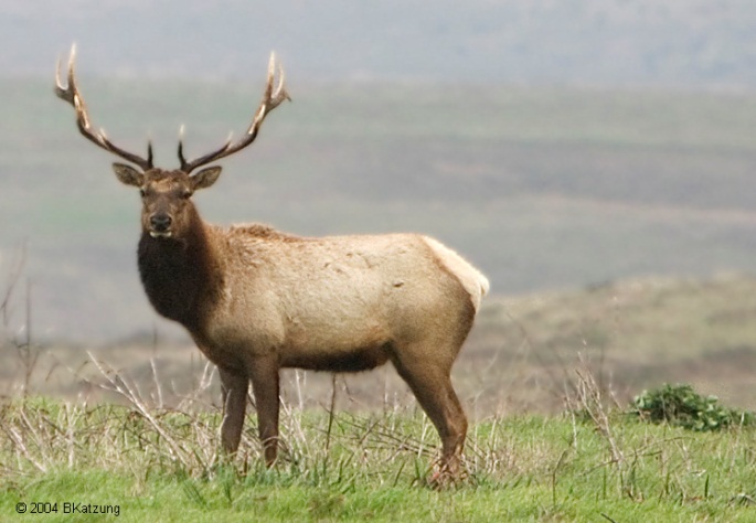
Tule elk (Cervus elaphus nannodes) at Point Reyes National Seashore
Recommended Publication:
Background (from that 2003 paper): The tule elk (Cervus elaphus nannodes) is a subspecies of C. elaphus and currently number some 3,700 individuals in several dozen populations in their North American native range. In 1978, 10 tule elk were released at the Point Reyes National Seashore (PRNS; California, USA) into what had for more than a century been a livestock range. An estimated 450 tule elk now [in 2003] live in a fenced 1,040-ha refuge at Tomales Point, an area of coastal prairie and coastal scrub in PRNS about 50 km north of San Francisco. Paratuberculosis in tule elk was first discovered in 1980 in three animals born at Point Reyes. The infection has subsequently been confirmed in additional tule elk and the organism has been isolated from dairy cattle, axis deer (Axis axis), and fallow deer (Dama dama) fecal samples in the Point Reyes area.
Abstract (with minor editing for consistency) : Forty-five adult tule elk (Cervus elaphus nannodes) in good physical condition were translocated from a population located at Point Reyes National Seashore, Marin County (California,USA), to a holding pen 6 mo prior to release in an unfenced region of the park. Because infection with Mycobacterium avium subsp. paratuberculosis (MAP) had been reported in the source population, the translocated elk underwent extensive ante-mortem testing using three Johne’s disease assays: enzyme linked immunosorbent assay (ELISA); agar gel immunodiffusion assay (AGID), and fecal culture. Isolation of MAP was made from fecal samples in six of 45 elk (13%). All AGID results were negative while ELISA results for 18 elk (40%) were considered elevated. Elevated ELISA results or MAP isolation from fecal samples were obtained for 22 of 45 elk (49%); these elk were euthanized and necropsied. Mycobacterium avium subsp. paratuberculosis was isolated from tissue in 10 of 22 euthanized elk (45%); of these 10 cases of confirmed infection, eight had elevated ELISA results (80%) and four were fecal culture positive (40%). One of 10 cases had histopathologic lesions consistent with MAP infection. Mycobacterium avium subsp. paratuberculosis was also isolated from tissue from one of eight fetuses sampled. The number of tule elk found to be infected was unexpected, both because of the continued overall health of the source herd and the normal clinical status of all study animals.
Comment: In all likelihood the elk acquired their MAP infection from cattle. Proving this is challenging, although the strains of MAP isolated from the elk are highly similar to those from cattle based on whole genome sequencing of two MAP strains from tule elk and several from cattle. The presence of paratuberculosis in this unique population of elk that is outgrowing the finite living space at PRNS presents challenges to those wanting to relocate some of elk from this population to new locations and to avoid depopulation by hunting or other means.
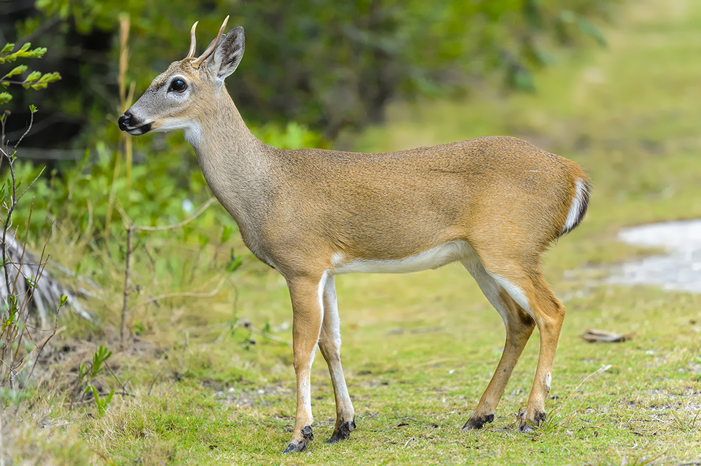
Key Deer (Odocoileus virginianus claviumin) Lower Florida Keys
Recommended Publication:
Background (from that 2008 paper): Johne’s disease was first identified in an endangered free-ranging Florida Key deer in 1996 at a private residence on Big Pine Key; a second case was confirmed 2 yr later at the same location (Quist et al., 2002). Based on a subsequent survey of repository serum and fecal samples and live capture, the prevalence of MAP infection was thought to be low in the Key deer population. However, from 2003 to 2004 five additional deer were diagnosed with Johne’s disease at the same residence and neighboring islands. These reports plus new findings in Johne’s disease research indicating that nonruminant wildlife are also susceptible to infection on heavily contaminated premises (Beard et al., 2001; Corn et al., 2005) raised the possibility that the infection prevalence had increased or was more extensive than previously thought. Additional concerns included illegal feeding of Key deer and the National Key Deer Refuge policy for translocation of deer to keys previously within the historic range of this species.
Abstract (with minor editing for nomenclature consistency): Johne’s disease, a fatal and contagious gastrointestinal infection caused by Mycobacterium avium subsp. paratuberculosis (MAP), was first diagnosed in an endangered Florida Key deer (Odocoileus virginianus clavium) in 1996 and later in six additional Key deer deaths from 1998 to 2004. We investigated the geographic distribution of MAP in the Lower Florida Keys from February 2005 through May 2006 via collection of blood and fecal pellets from 51 live-captured deer, collection of 550 fecal samples from the ground, and by necropsies of 90 carcasses. Tissue and fecal samples also were submitted from 30 raccoons (Procyon lotor), three feral cats (Felis catus), an opossum (Didelphis virginiana), and a Lower Keys marsh rabbit (Sylvilagus palustris hefneri). Mycobacterium avium subsp. paratuberculosis was identified in 23 Key deer fecal samples collected from the ground, tissue samples from two clinically ill Key deer, and from the mesenteric lymph node of a raccoon. The results of this study indicate that MAP persists in the Key deer population and environment at a low prevalence, but its distribution currently is limited to a relatively small geographic area within the range of Key deer.
Fallow Deer (Dama dama) in Spain

Recommended publication:
Background (from that 2008 paper):The Regional Hunting Reserve of El Sueve (Principado de Asturias, in northern Spain; 43⁰ 15’N, 5⁰ 15’E) is a mountainous area of 8,300 ha, 5 km from the seashore. Fallow deer were introduced in 1960. Since then the population has increased so that population reduction was necessary for Reserve management. The population of fallow deer was estimated to be approximately 1,000 individuals in 1999. During summer there were about 1,200 cattle, 550 horses, and 850 sheep and goats that shared pastures and water holes with the deer.
Abstract (with minor editing for nomenclature consistency): Paratuberculosis was diagnosed in a population of approximately 1,000 free-ranging fallow deer (Dama dama) sampled from 1997–98 in the Regional Hunting Reserve of El Sueve (Asturias, Spain). Five of eight animals observed with diarrhea were diagnosed as having paratuberculosis on the basis of gross lesions at postmortem examination and histopathology. In two deer, Mycobacterium avium subsp. paratuberculosis was cultured and identified by polymerase chain reaction. Indirect enzyme-linked immunosorbent assay and immunodiffusion tests were used to evaluate sera from 33 adult deer from this population. All fallow deer tested were seronegative.
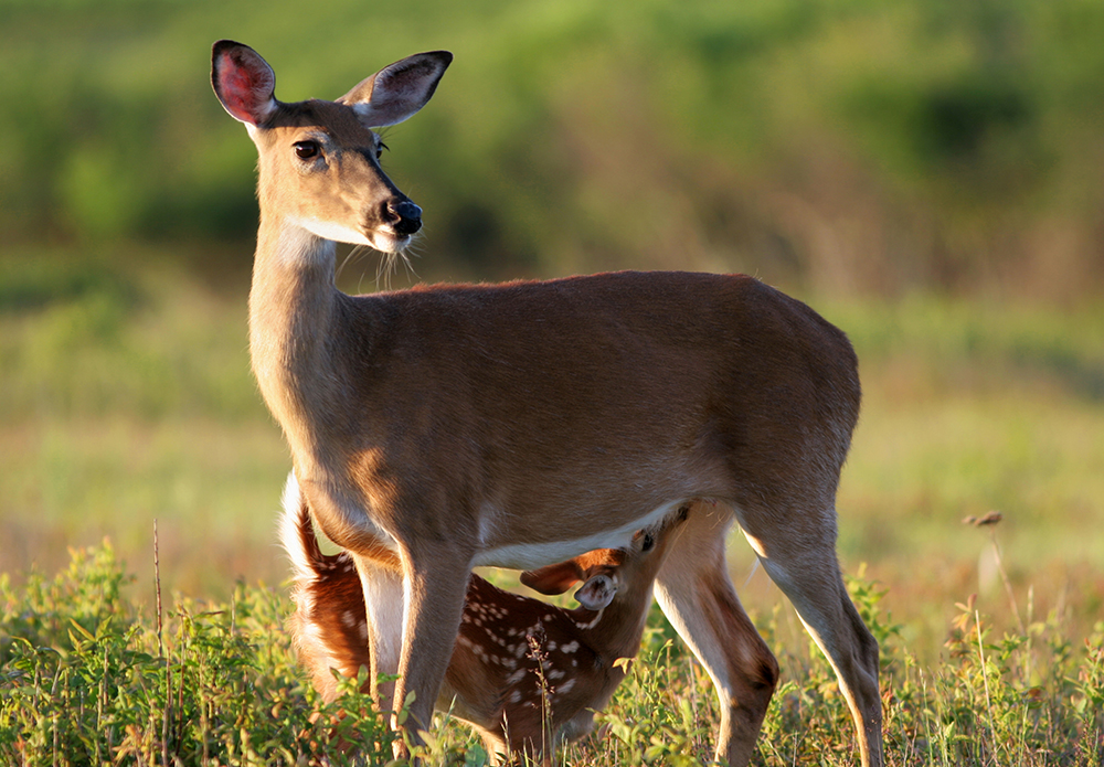
White-tailed Deer (Odocoileus virginianus)
Recommended Publication:
Background (from that 2009 paper): In October 2006, following several reports of sick white-tailed deer, a 2-yr-old,male deer from Fauquier County, USA, was reported to the Virginia Department of Game and Inland Fisheries (VDGIF) field staff as being recumbent and reluctant to move. Notable clinical signs included emaciation and abundant dried fecal material around the perineum and hind legs. The deer was euthanized by cervical gunshot, which is an agency-approved method, and a field necropsy was performed. Gross necropsy findings included markedly poor body condition with prominent ribs and vertebrae as well as minimal body fat. The gastrointestinal tract (small intestines, cecum, spiral colon, colon, and rectum) contained copious amounts of brown-green malodorous liquid feces. The serosal surface of the gastrointestinal tract was erythematous with congested and tortuous blood vessels. Additionally, areas of petechial hemorrhage were present on the mucosal surface of various sections of the intestinal tract. The mesenteric lymph nodes were markedly enlarged, and the liver contained several small, white foci within the parenchyma.
Abstract (with minor editing for nomenclature consistency): Johne’s disease (paratuberculosis) was diagnosed in a 2-yr-old, male, free-ranging white-tailed deer (Odocoileus virginianus) from Fauquier County, Virginia, USA, based on histopathology and culture for Mycobacterium avium subspecies paratuberculosis. Clinical and pathologic findings included emaciation; loss of body fat; chronic diarrhea; severe, chronic, diffuse granulomatous colitis with intrahistiocytic acid-fast bacilli; moderate, chronic granulomatous lymphadenitis with intrahistiocytic acid-fast bacilli; as well as moderate chronic, multifocal, lymphoplasmacytic hepatitis. These findings are consistent with previous reports of Johne’s disease in cervids. Subsequent targeted surveillance of 10 emaciated deer with diarrhea, as well as sampling of 72 asymptomatic deer for M. avium subsp. paratuberculosis using culture of multiple tissue types, as well as serology using an enzyme-linked immunosorbent assay (ELISA) optimized for cervid antibody detection, did not reveal any additional cases of infection in this geographic region. To date, this appears to be an isolated case of Johne’s disease in a free-ranging white-tailed deer, and infection with the causative agent for Johne’s disease appears to be an infrequent occurrence in deer from this region. The origin of infection was most likely domestic ruminants. This is the first report of clinical Johne’s disease in a free-ranging white-tailed deer outside of the Florida Keys, USA. Stressors, such as high deer population density and low selenium levels, may have contributed to the development of clinical disease in this case and warrant further investigation.
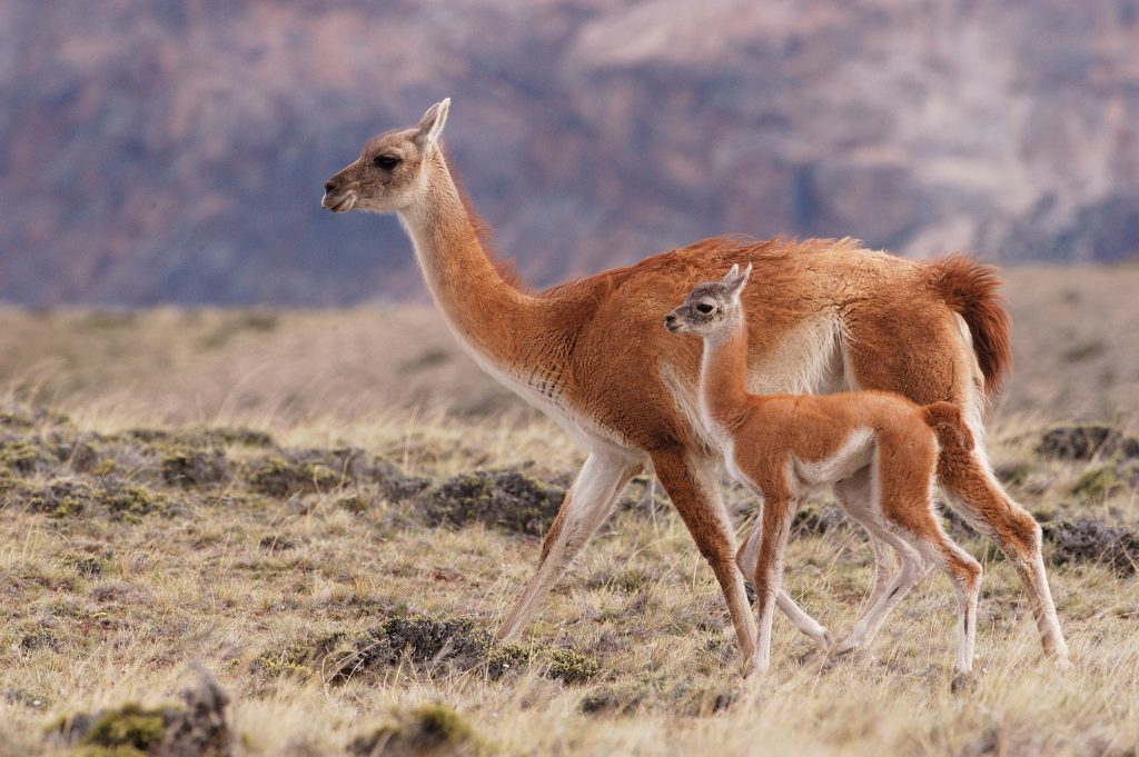
Guanaco (Lama guanicoe)
Recommended Publication:
Background (from that 2009 paper): The guanaco (Lama guanicoe) is the largest South American camelid, naturally distributed between 8⁰S latitude in Peru and 55⁰S S on Tierra del Fuego Island (Patagonian Region) at altitudes from the sea level to 4,600 m. The guanaco population in Chile is about 86,000 and most are concentrated in the Patagonian Region. It is estimated that approximately 45,000 guanacos live in the southern part of Tierra del Fuego Island. The guanaco is the only wild ungulate species widely distributed across the Patagonian steppe and shares grazing land with approximately one million sheep and about 20,000 cattle (Chile, 2007). It is considered a broad-range herbivore, eating grass on pastures or bush according to seasonal food availability. Guanaco prefer open habitats occupying steppe and prairies, although they also make use of forest habitat. Its behavior can be described as seasonally territorial but during harsh and changing environmental conditions social group composition and size may change (Puig and Videla, 1995). In wild guanaco populations three different basic structures are recognized: family groups, groups of young males, and solitary individuals. There is no published information on MAP in this species.
In order to control guanaco overpopulation on the Chilean island of Tierra del Fuego, and in order to protect the land from the deleterious effect of these animals on the native forest, the Regional Agriculture and Livestock Department of the Ministry of Agriculture of Chile authorized a supervised yearly hunting quota of 2,000 animals, respecting preservation of the species. The aims of this study were to test these animals for MAP.
Abstract: The aim of this study was to search for Mycobacterium avium subsp. paratuberculosis (MAP) infection in a free-ranging wild animal species in a region where Johne’s disease has yet to be reported and to classify Map isolates using a genomic typing method. Fecal samples were obtained from 501 wild guanacos (Lama guanicoe) from Tierra del Fuego Island, Chile, in August 2006. Samples were cultured using Herrold’s egg yolk medium with and without mycobactin J. After 9 mo. of incubation, suspected MAP colonies showing mycobactin dependence were confirmed by real-time polymerase chain reaction (PCR) based on IS900 and F57. Isolates were further tested using IS1311 PCR with restriction endonuclease analysis in order to type the guanaco MAP strains. Twenty-one of 501 (4.2%) animals were fecal culture-positive for MAP; identity was confirmed by real-time PCR and isolates were classified as cattle-type. Most culture-positive animals were located in four contiguous geographic areas, and the infection was most commonly found among adult animals. Prevalence was higher in females (5.9%) than males (3.1%) but the difference was not statistically significant. This represents the first isolation of MAP from a free-ranging wildlife species in Chile. It expands the geographic range of paratuberculosis and the diversity of wildlife species that can become infected with MAP.
Reindeer (Rangifer tarandus) also known as the caribou in North America
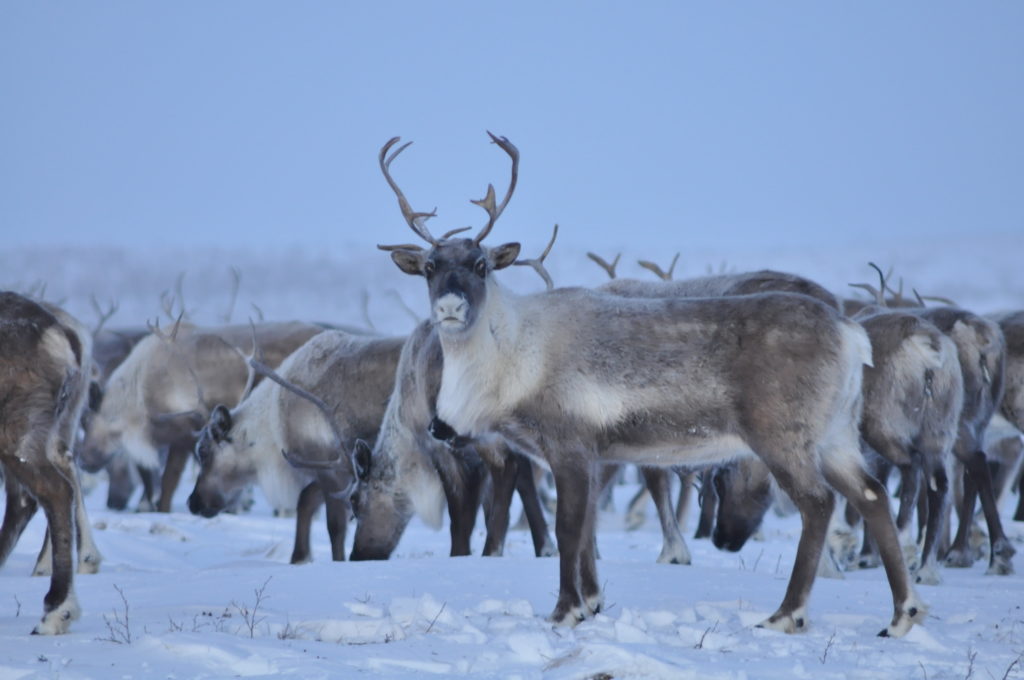
Background (excerpt from publication): Caribou and reindeer (R. tarandus spp.) are keystone species in the Arctic and Subarctic and central to the cultural, socioeconomic, and physical well-being of many circumarctic peoples. The health and sustainability of Rangifer is thus of high priority to circumpolar nations. Johne’s disease described in semi-domestic reindeer herds has an acute onset and results in a high mortality rate (Katic, 1961). In this species, diarrhea is generally absent and weight loss is the typical sign of disease (Poddoubski, 1957). Mycobacterium avium subspecies paratuberculosis antibody-positive semi-domestic reindeer (Tryland et al., 2004) and wild caribou (Johnson et al., 2010) are reported, and MAP has been isolated from tissues of captive reindeer in poor body condition in Yukon, Canada (Merchant, pers. comm.). Our goal was to determine if MAP occurs in wild caribou in northern Canada and Greenland and establish baseline data regarding its distribution among these herds.
Abstract: Mycobacterium avium subspecies paratuberculosis (MAP) is a common pathogen in domestic ruminants that causes granulomatous inflammation of the small intestine leading to emaciation and wasting. Clinical disease (Johne’s disease) is also reported for several wild ruminant species. Between 2007 and 2009 we collected 561 fecal samples from caribou (Rangifer tarandus ssp.) representing 10 herds of migratory caribou, two herds of caribou from Greenland, and three populations of boreal woodland caribou. Feces were tested for MAP by bacterial culture and PCR targeting the IS900 insertion sequence. In total, 31 samples from eight different populations representing all three ecotypes were found positive for MAP by PCR, with one sample from the Rivière-aux-Feuilles herd also being culture positive for the type II (cattle) strain. The proportion of positive animals was particularly high in the Akia-Maniitsoq herd in Greenland, and Rivière-aux-Feuilles and Rivière-George herds in northeastern Canada (23.4, 11.5, and 10.0%, respectively). Our results indicate that MAP is present in several caribou herds of different ecotypes in northern Canada and Greenland and that MAP circulates within wildlife populations that do not have ongoing contact with domestic livestock. The epidemiology, pathogenicity, and effects on the health of caribou in northern ecosystems remain unknown.
Huemel deer (Hippocamelus bisulcus) of Southern Patagonian [endangered]
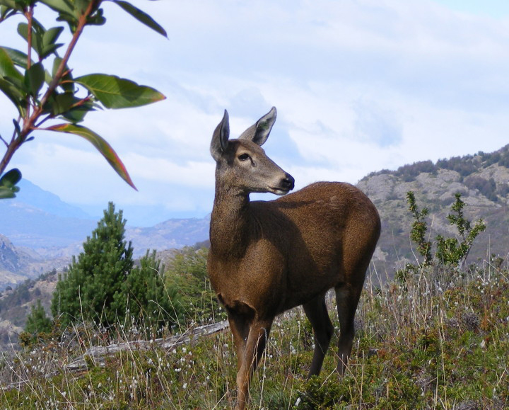
Background (excerpt from publication): The International Union for the Conservation of Nature (IUCN) estimates that over 24% of the world’s extant mammals are currently threatened with extinction, yet infectious diseases have only been listed as a major threat for a small fraction (1.1%). It is likely that diseases are underrepresented as a contributing threat to wildlife extinction, especially given that less than half (39%) of critically endangered and endangered artiodactyls, carnivores, and primates from the 2006 IUCN Red List have any published records of pathogens from their wild populations (Pedersen et al 2007). Recently, evidence linking huemul (Hippocamelus bisulcus), an endemic and endangered deer species from southern Chile and Argentina (Corti et al 2010), with Mycobacterium avium subsp. paratuberculosis (MAP) infection has been reported (Salgado et al 2017). MAP is the causative agent of Johne’s disease, infection mostly affecting domestic ruminants, but it has been shown to pose a threat also to wildlife (Manning and Collins 2001, Salgado et al 2015). However, the pathogen has also been isolated from non-ruminants species and even from humans, associating it with Crohn’s disease (Chiodini et al 2012). Although huemul inhabits remote areas with limited contact with domestic animals/livestock and exists at low population density, the presence of pathogens like MAP may be interpreted as an indicator of spill-over infections from domestic animals.
Abstract: In a huemul (Hippocamelus bisulcus) population sympatric with cattle, we found evidence of Mycobacterium avium subsp. paratuberculosis (MAP) infection. Three huemul faecal pellet samples and two cow pats were collected and cultured for MAP presence. DNA was then extracted for PCR analysis of all signal-positive cultures. To assess whether MAP isolates obtained from huemul faeces were associated with typical MAP isolated from livestock, positive confirmed culture samples were sub-typed using a combination of five Mycobacterial Interspersed Repetitive Unit-Variable Number Tandem Repeat Analysis and one Short Sequence Repeat analysis markers. All faecal samples from both species were MAP positive. One huemul presented a different bacteria profile genotype not described before, suggesting that huemul and cattle in Patagonia could carry a unique MAP strain.