

G.V. Sandoval and colleagues in Argentina reported on the rate of intrauterine MAP transmission in pregnant Saanen goats.
Their findings were reported in a recent issue of the journal Veterinary Microbiology.
ABSTRACT
Paratuberculosis (PTBC) is a chronic granulomatous enteritis of ruminants caused by Mycobacterium avium subsp. paratuberculosis (Map). Although PTBC intrauterine transmission has been described in cattle, sheep, and deer, data in dairy goats are limited. The present study investigated intrauterine Map infection in 32 pregnant Saanen goats from a herd with high PTBC prevalence in Jujuy, Argentina. Maternal and fetal tissues were analyzed using a range of methods, including bacteriological culture, IS900 PCR, histopathology, and serology. All goats were positive to the intradermal test, and 75% showed histopathological lesions consistent with PTBC. Map was isolated from 10 of 45 fetuses. Granulomatous lesions were observed in two cases, but no acid-fast bacilli were detected. Furthermore, fetuses from culture-positive dams were 7.8 times more likely to be infected than those from culture-negative ones. This suggests that the maternal bacterial load is a key predictor of fetal infection. These findings represent the first evidence of intrauterine transmission of Map in Saanen goats, even in subclinical cases, indicating that vertical transmission may be more common in this species than previously assumed. This has significant implications for PTBC control programs in small ruminants.
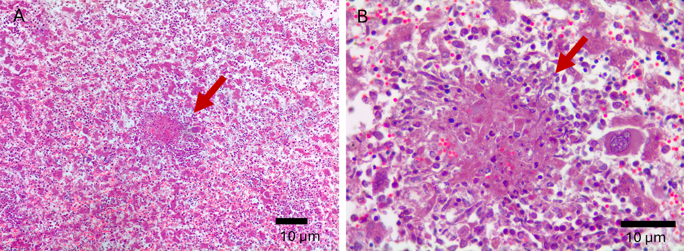
Liver granulomas from MAP-infected female goats indicating they had a disseminated infection. H&E stain. A. 10x magnification B. 40x magnification. Sandoval et al. Veterinary Microbiology 315 (2026) 110954.
COMMENTS
This intriguing study that verifies that MAP infections can pass from mother (dam) to off-spring before birth, i.e., in utero. The best predictors of whether in utero transmission would occur were culture of maternal tissues collected at necropsy and PCR on these same tissues. Interestingly, ELISA on the dams did not predict the likelihood fetal infection, i.e. there was no statistical association between the ELISA-positive status of the pregnant goat and the MAP infection status of its fetus.
Why this study might not represent all goat herd situations:
- This study was done on a single heavily MAP-infected herd of Saanen goats.
- The herd had a very high MAP infection rate, estimated to be 23%. This means that the female goats in the study were likely exposed to very high numbers of MAP causing more rapid infection progression. These females themselves may have become MAP-infected in utero.
Take home messages for herd owners working to control or eradicate Johne’s disease:
- PCR is preferable to ELISA for herd testing, especially for breeders.
- It is best to not keep as herd replacements any kids born to PCR-positive dams.
- Owners cannot simply trust their eyes to diagnose MAP-infected goats. All dams in this study were in good body condition (body condition score over 3), but 14 of 32 had advanced MAP infections based on histopathology. Weight loss and diarrhea do not happen until very late in the course of a MAP infection in goats. Thus, goat owners must rely on PCR on fecal samples to detect MAP-infected goats, although this study did not report fecal PCR test results on live goats but instead relied on PCR on tissue samples collected at necropsy.