
This question was recently submitted to our website’s “Ask and Expert” feature:
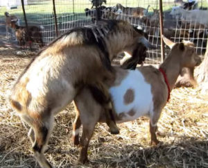 “I have a goat buck who has tested positive for Johne’s. If the diagnosis is confirmed, is it OK to collect his semen to store and use for AI purposes? Or can it spread the disease? Should I wait a couple of years before using it to be sure that it is safe?”
How would you answer this goat owner?
There are many reports that bulls (bovine) infected with MAP have the organism in their semen and accessory sex glands. Research publication examples include the following:
- AB Larsen. Mycobacterium paratuberculosis in reproductive organs and semen of bulls. Am. J. Vet.Res. 31:255, 1970.
- WY Ayele. Distribution of Mycobacterium subsp. paratuberculosis in organs of naturally infected bull-calves and breeding bulls. Vet. Microbiol. 103:209, 2004.
- JL Kohl. Consecutive excretion of Mycobacterium avium subspecies paratuberculosis in semen of a breeding bull compared to the distribution in feces, tissue and blood by IS900 and F57 quantitative real-time PCR and culture examinations. J. Vet. Med. Sci. 72:1283, 2010.
- P Münster. A Longitudinal study to characterize the distribution patterns of Mycobacterium avium ssp. paratuberculosis in semen, blood and faeces of a naturally infected bull by IS900 semi‐nested and quantitative real‐time PCR. Transboundary and Emerging Diseases. 60:175, 2013.
While it seems that there is consensus that MAP is found in the semen of infected bulls, a search of scientific literature indicates that only Dr. Richard Merkal tackled the question of what the consequences are for the cow inseminated with MAP-contaminated semen (RS Merkal. Intrauterine inoculation of Mycobacterium paratuberculosis into guinea pigs and cattle. Am. J. Vet. Res. 43:676, 1982).
The study was small (3 cattle) and used a large intrauterine inoculum, 5×10⁸ MAP, and necropsied the cattle up to 4 weeks post-inoculation. MAP was recovered from the body and horns of the uterus of inoculated cattle at 1, 2, 3, and 7 days post-inoculation and from a pelvic lymph node of one cow necropsied 2 weeks post-inoculation but not from tissues of cattle inoculated at 3 and 4 weeks post-inoculation. [The article is provided in full here because of its age and uniqueness. Apologies for the poor photocopy quality.]
Comment: In my answer to the goat owner’s question I explained that I was not aware of any research that directly addressed this question in goats but that I did have information on cattle (some cited above). Based on this, I suggested that the semen itself was probably of minimal concern: best summarized as “wrong end & wrong age”. I also explained that the feces of the infected buck were of greater concern.
Although Merkal’s study raises some concerns, the uterus is the wrong end (oral being the preferred route of infection by MAP) and only adult (post-puberty) animals are inseminated and as such are more resistant to MAP infection than young animals, i.e. wrong age.
However, I think this highlights a knowledge gap in our understanding about Johne’s disease. This has major significance for the artificial insemination (AI) industry and for countries like Sweden that are free of MAP infections in cattle and risk importation of the infection. And, we obviously know even less about MAP in goat bucks.