
Research Report
MV Palmer and colleagues from the U.S. National Veterinary Services Laboratory (NVSL) reported a detailed investigation of Johne’s disease in a captive white-tailed deer herd. The article appears in the Journal of Veterinary Diagnostic Investigation 31(6):844–851, 2019.
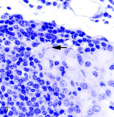
So few MAP but so much inflammation!
Abstract
Paratuberculosis (Johne’s disease) is caused by Mycobacterium avium ssp. paratuberculosis (MAP), and affects both domestic and wild ruminants, including cattle, goats, sheep, and deer. In cattle, most infections occur during calfhood followed by a prolonged incubation period of 1–2 y or more before cows shed culturable numbers of MAP bacilli in their feces. As disease progresses, infected animals develop protein-losing enteropathy, intractable diarrhea, and weight loss. In a cohort of 32 clinically normal deer from a herd with a history of periodic clinical paratuberculosis, we found that subclinical infection was characterized by high rates of infection, common involvement of mesenteric lymph nodes, minimal lesion formation, few intralesional acid-fast bacilli, and low-level fecal shedding of MAP. The characteristics of subclinical paratuberculosis in white-tailed deer resemble those of cattle and red deer, although microscopic lesions were less common in subclinical deer than reported for subclinical cattle, and we did not see necrotizing granulomas as described in subclinical red deer and elk.
Comment: This report describes a very thorough investigation with repeated sampling and extensive examination of tissues after necropsy. The photomicrographs are particularly nice and highlight how hard it is to find MAP in tissue sections, even with special stains. Unfortunately, this article is not Open Access.