
JOHNE’S IN WHITE-TAILED DEER
2019-12-02 17:25:28Research Report
MV Palmer and colleagues from the U.S. National Veterinary Services Laboratory (NVSL) reported a detailed investigation of Johne’s disease in a captive white-tailed deer herd. The article appears in the Journal of Veterinary Diagnostic Investigation 31(6):844–851, 2019.
So few MAP but so much inflammation!
Abstract
Paratuberculosis (Johne’s disease) is caused by Mycobacterium avium ssp. paratuberculosis (MAP), and affects both domestic and wild ruminants, including cattle, goats, sheep, and deer. In cattle, most infections occur during calfhood followed by a prolonged incubation period of 1–2 y or more before cows shed culturable numbers of MAP bacilli in their feces. As disease progresses, infected animals develop protein-losing enteropathy, intractable diarrhea, and weight loss. In a cohort of 32 clinically normal deer from a herd with a history of periodic clinical paratuberculosis, we found that subclinical infection was characterized by high rates of infection, common involvement of mesenteric lymph nodes, minimal lesion formation, few intralesional acid-fast bacilli, and low-level fecal shedding of MAP. The characteristics of subclinical paratuberculosis in white-tailed deer resemble those of cattle and red deer, although microscopic lesions were less common in subclinical deer than reported for subclinical cattle, and we did not see necrotizing granulomas as described in subclinical red deer and elk.
Comment: This report describes a very thorough investigation with repeated sampling and extensive examination of tissues after necropsy. The photomicrographs are particularly nice and highlight how hard it is to find MAP in tissue sections, even with special stains. Unfortunately, this article is not Open Access.
MULTIPLEX MAP PCR AT THE WVDL
2019-11-25 14:30:36Announcement from the Wisconsin Veterinary Diagnostic Laboratory (WVDL)
 The WVDL has completed the validation of new multiplex real-time PCR assay for the detection of Mycobacterium avium ss. paratuberculosis (MAP) in accordance with the American Association of Veterinary Laboratory Diagnosticians (AAVLD) guidelines. WVDL has improved the specificity and sensitivity of the assay for all species routinely tested including for bovine, caprine, ovine and exotic samples. This multiplex real-time PCR assay uses three targets, which minimizes the possibility of cross-reaction with other Mycobacterium avium complex members thereby increasing MAP detection specificity. The validation data showed that only a very a small number of exotic samples (< 0.01%) cannot be resolved using the multiplex PCR. In those situations, the WVDL reports the MAP PCR result as “undetermined”, with the additional recommendation of retesting the animal within the next 6 months. If you have any questions, please feel free to contact the Microbiology Molecular Section at the WVDL by phone at 608-262-5432, by email at info@wvdl.wisc.edu or at our website.
Comment: Multiplex PCR assays are not new, but this is among the first reports that the assay has been validated and made routine in a veterinary diagnostic laboratory.
I have been involved in two situations where a commonly used commercial MAP PCR kit reported a positive result on zoological ruminants only to discover after more extensive testing that the animals had a strain of M. avium subsp. avium in their fecal sample that caused a false-positive MAP PCR kit result. The 3-target multiplex has been proven to avoid this potentially costly mistake. When a MAP diagnosis by conventional real-time PCR kit has large economic consequences, it is advisable to “get a second opinion” using this multiplex PCR assay, or simply start with this multiplex assay given that the cost is comparable to what most labs charge using the single genetic target PCR kits for MAP.
____________
For the very interested, here are some research reports on multiplex PCR assays for MAP:
- DV Cousins et al. Mycobacteria distinct from Mycobacterium avium subsp. paratuberculosis isolated from the faeces of ruminants possess IS 900-like sequences detectable by IS 900 polymerase chain reaction: implications for diagnosis. Molecular and Cellular Probes 13(6):431-442, 1999.
- T Tasara et al. Development and evaluation of a Mycobacterium avium subspecies paratuberculosis (MAP) specific multiplex assay. International Journal of Food Microbiology 104(3):279-287, 2005.
- S Rajeev. Evaluation of multiple genomic targets for identification and confirmation of Mycobacterium avium subsp. paratuberculosis isolates using real-time PCR. Veterinary Microbiology 105(3-4):215-221, 2005.
- D Herthnek and G Bölske. New PCR systems to confirm real-time PCR detection of Mycobacterium avium subsp. paratuberculosis. BMC Microbiology 6: article no. 87, 2006.
- SJ Shin et al. Efficient differentiation of Mycobacterium avium complex species and subspecies by use of five-target multiplex PCR. Journal of Clinical Microbiology 48(11):4057-4062, 2010.
AORTIC CALCIFICATION WITH JOHNE’S DISEASE
2019-11-18 00:46:45Everyday there is something interesting I learn about Johne's disease and it comes from diverse sources. This story is about -
comparative pathology
and
collaborative learning
The email below was received last week from Dr. Fritz Schumann, a Canadian veterinarian working in Saskatoon (see map):

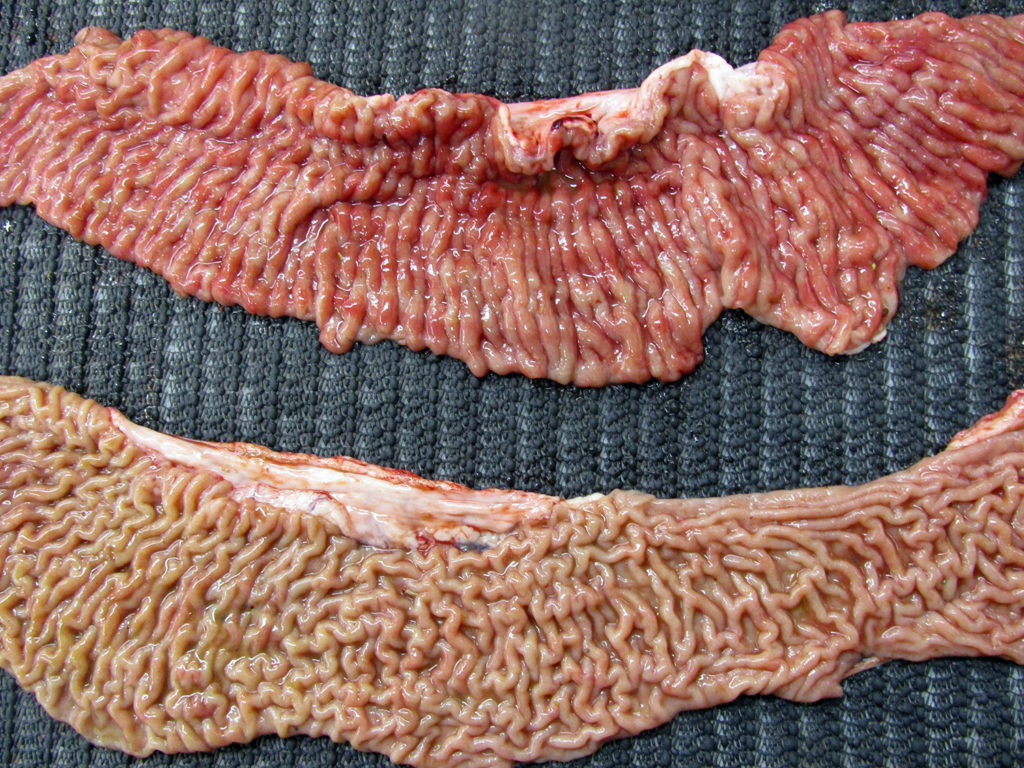
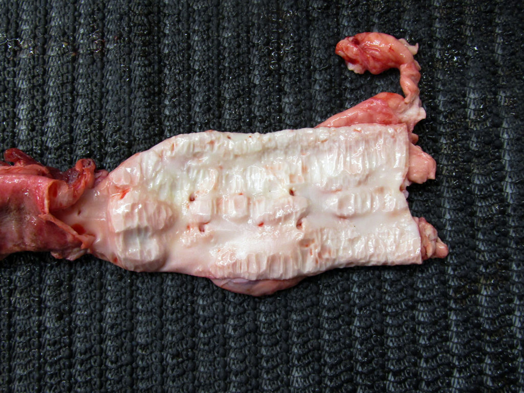
He wrote: I am vet in Saskatoon. I had this 2 year and 9-month-old Black Angus cow which I euthanized as she had Johne’s. Positive ELISA in blood and PCR positive on feces. I euthanized her and am sending two pictures. The first is her ileum and jejunum. The second picture is the aorta, looks like calcification from the aortic valve all the way to the pelvis then I stopped looking. The aorta felt like there is no elastic tissue left and paper thin. My question is why does aorta change like that with Johne’s? This is the worst aorta I have seen. Thank you very much for reading my email and considering my request for an explanation.
THANK YOU DR. SCHUMANN! He granted me permission to show his exceptional pictures taken at the Western College of Veterinary Medicine. The first shows the classic thickened ileum (top) and the jejunum for comparison (bottom).
This second photo shows the striking degree of calcification of the aorta.
I made inquiries with colleagues and this reply came from the Minnesota Veterinary Diagnostic Laboratory pathologists. Below is their quick and informative reply. Big thanks! to these pathologists.
Provided here is an excerpt from a paper that was published in JVDI on blastomycosis in a llama (J Vet Diagn Invest. 2018 Jul; 30(4): 576–579). It provides some thoughts on possible pathogenesis:
"As a component of chronic disease, monocytes and macrophages can generate and release tumor necrosis factor–alpha (TNFα) that drives mineralization, and macrophages may accumulate basic calcium phosphate that can locally increase cytokine production (TNFα, interleukin [IL]-1, and IL-8) that further drives mineralization and stimulates endothelial cells to differentiate toward osteoblasts (20). In dromedary camelids and cattle with Johne’s disease, which exhibits a predominantly granulomatous cellular response, proinflammatory cytokines (IL-1α, IL-1β, IL-6, IL-10, interferon-γ, and TNF-α), acute-phase and oxidative-stress proteins are significantly increased (9). Hypercalcemia has been reported in dogs with granulomatous diseases, including blastomycosis, and is likely associated with the conversion of calcifediol (25-hydroxyvitamin D) to calcitriol (1,25-dihydroxyvitamin D) by activated macrophages (8,14)."
Citations mentioned in this 2018 JVDI excerpt:
- #8. Dow SW, et al. Hypercalcemia associated with blastomycosis in dogs. J Am Vet Med Assoc 1986;188:706–709.
- #9. El-Deeb W, et al. Clinico-biochemical investigation of paratuberculosis of dromedary camels in Saudi Arabia: proinflammatory cytokines, acute phase proteins and oxidative stress biomarkers. Pak Vet J 2014:34;484–488.
- #14. Meuten D. Parathyroid glands and calcium and phosphorus metabolic pathology. In: Thrall MA, et al., eds. Veterinary Hematology and Clinical Chemistry. 2nd ed. Ames, IA: WileyBlackwell, 2012:545–568.
- #20. Shantsila E, Lip GY. Systemic inflammation as a driver of vascular calcification: a proof of concept. J Intern Med 2009;266:453–456.
I am also providing a link to the excellent 1978 article by Dr. Claus Buergelt and colleagues on the pathology of bovine paratuberculosis which mentions aortic calcification.
______________
Comment: This report is a perfect example of how an inquisitive veterinary practitioner requested a necropsy on a cow with Johne's disease, how veterinary pathologists documented some novel pathology, how the practitioner reached out via the internet to learn more, and how personal and internet connections helped everyone reading this post to know more about the pathobiology of Johne's disease. In many ways it mirrors the how the first report of Johne's disease came to be.
______________
And.....for the keenly interested, here are some additional related references with links. This is not an exhaustive list. Sorry if I missed your favorite publication:
- US Sorge et al. Cow-level association between serum 25-hydroxyvitamin D concentration and Mycobacterium avium subspecies paratuberculosis antibody seropositivity: A pilot study. Journal of Dairy Science. 96(2):1030-1037, 2013.
- JR Stabel et al. Dietary calcium modulates Mycobacterium paratuberculosis infection in beige mice. Veterinary Immunology and Immunopathology, 66(3–4): 377-390, 1998.
- RW Jayalath et al. Aortic calcification. European Journal of Vascular and Endovascular Surgery. 30(5):476-488, 2005.
- N Niederhoffer et al. Aortic calcification produced by vitamin D3 plus nicotine. Journal of Vascular Research. 34(5):386-398. 1997.
MAP IN DAIRY GOATS – INDIA
2019-11-15 14:07:49 Manju Singh and colleagues reported on detection of MAP infections in dairy goats in India using 6 tests to screen 465 raw milk samples. The lead author is from the Department of Biotechnology, Institute of Applied Sciences & Humanities, GLA University, Mathura, Uttar Pradesh, India and AIMT & AIB, Amity University Rajasthan, Jaipur, India. The article appears in the journal Comparative Immunology, Microbiology and Infectious Diseases, volume 64, pages 53-60, June 2019. Amity University Rajasthan in Jaipur will be the site of the 16th International Colloquium on Paratuberculosis, October 17-21, 2022.
Abstract
Johne's disease, caused by Mycobacterium avium subspecies paratuberculosis is endemic in the domestic livestock population, still it is not priority for control in the country. First time we used 'multiple assays’ for screening raw milk of 465 goats (farm/farmer's herds) to estimate bio-load and bio-type profile of bacilli. Each sample was screened by six tests and compared their sensitivity and specificity. Of 465 raw milk samples screened, bio-load of bacilli was 65.3% by six assays. Assay-wise bio-load was 49.4 and 62.7% in antigen and antibody detection tests, respectively. Bio-load was 48.8, 46.6, and 13.9% in Indirect Fluorescent Antibody Test (i_FAT), microscopy and IS900 PCR and 39.1, 57.4 and 55.6% in Indirect Enzyme Linked Immuno Sorbant Assay (i_ELISA), Dot Enzyme Linked Immuno Sorbant Assay (d_ELISA) and Latex Agglutination Test (LAT), respectively. Dot-ELISA was most sensitive followed by LAT, i_FAT, microscopy and i_ELISA. Milk DNA samples positive in IS900 PCR on bio-typing using IS1311 PCR_ Restriction Enzyme Analysis (IS1311 PCR_REA) revealed, 72.3% (47/65) were 'Indian Bison Type'. Milk was easy to collect sample and first time we used 'whole milk' as 'test sample' without centrifugation. High bio-load of MAP in milk underlined need for urgent control of disease in lactating goatherds. Bacilli was important 'Milk born' infection and on the basis of sensitivity, specificity, resources and requirements, of the ‘six assays’ most appropriate assay/s (single or in combination) can be chosen for the screening and diagnosis of Johne’s disease in lactating goatherds using whole milk as sample.
Conclusion (from publication)
Bio-load of MAP was high in goats on the basis of screening of raw milk using six assays. Study compared efficacy of six assays and identified better diagnostic test / test combination ('testing strategy') for detecting presence of MAP in goat milk / goats. Study established the role of milk as carrier of MAP to newborn animals and human population. Study showed that MAP was the major, 'Milk born pathogen'. Thereby, helping in perpetuation of MAP infection generations to generations in animals and from animals to human population. The choice of test for screening of a population depends on the purpose and resources available. LAT, d_ELISA and microscopy had potential to be good screening assays for screening of 'raw milk'. Whereas, i_FAT and i_ELISA could be used as confirmatory assays. Milk was convenient and good clinical sample for estimating bio-load of MAP in lactating animals.
Comment: The term bio-load, used throughout this publication, seems to equate to rate of positivity for each of the 6 diagnostic tests evaluated, rather than the quantity of MAP in the samples. Since there was no goat population proven to be free of MAP infections, it is difficult to assess the true specificity of each of the 6 assays. It is noteworthy, however, that 65 (13.9%) of samples were IS900 PCR-positive and that the majority (72.3%, 47/65) were bio-typed as ‘Indian Bison Type’, the remainder could not be bio-typed.
15TH INTERNATIONAL COLLOQUIUM ON PARATUBERCULOSIS
2019-11-08 14:57:58![]() Only 22 days left to submit your abstract!
Only 22 days left to submit your abstract!
Abstract submission for the 15th International Colloquium on Paratuberculosis to be held in Dublin, Ireland on June 14th to 18th has opened at this link and will remain open until December 1st. Hope that you will submit an abstract! Looking forward to a great conference next Summer.
Complete information about the upcoming 15-ICP can be found here.
HEALTHY COWS - HEALTHY KIDS
2019-11-03 17:17:40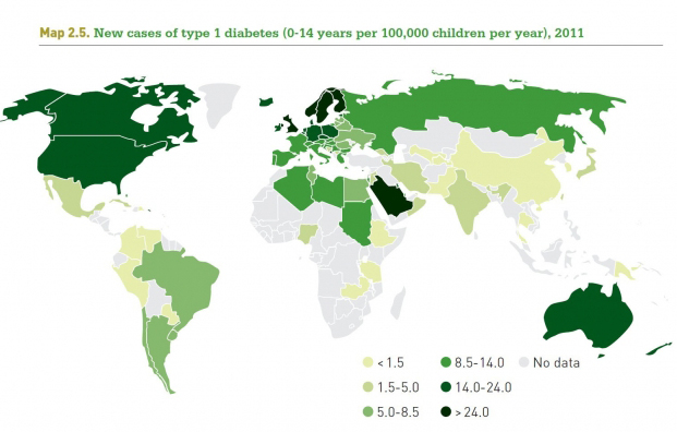 In the journal Microorganisms, Dr. C.T. Dow and L.A. Sechi present a hypothesis paper titled: Cows Get Crohn’s Disease and They’re Giving Us Diabetes. This 10-page article with 111 references was published 17-OCT-2019 and is an Open Access article and is a very worthy read.
Abstract
Increasingly, Johne’s disease of ruminants and human Crohn’s disease are regarded as the same infectious disease: paratuberculosis. Mycobacterium avium ss. paratuberculosis (MAP) is the cause of Johne’s and is the most commonly linked infectious cause of Crohn’s disease. Humans are broadly exposed to MAP in dairy products and in the environment. MAP has been found within granulomas such as Crohn’s disease and can stimulate autoantibodies in diseases such as type 1 diabetes (T1D) and Hashimoto’s thyroiditis. Moreover, beyond Crohn’s and T1D, MAP is increasingly associated with a host of autoimmune diseases. This article suggests near equivalency between paucibacillary Johne’s disease of ruminant animals and human Crohn’s disease and implicates MAP zoonosis beyond Crohn’s disease to include T1D.
Facts to know:
Worldwide, the incidence of T1D is increasing. Over the last 25 years, the incidence went up by over 12%. It was 10.9 per 100,000 among children aged 0-14 years in 1990 and rose to 22.5 per 100,000 in 2009. (Type 1 Diabetes Statistics)
The current prevailing paradigm on the etiology of T1D hypothesizes that environmentally triggered autoimmune destruction of pancreatic beta cells occurs against the background of genetic risk. (Endocrinol Metab Clin North Am. 2010 Sep; 39(3): 481–497).
Comment:
Healthy food comes from healthy animals. It is a reasonable consumer expectation that foods do not harbor zoonotic pathogens, and MAP has earned the zoonotic pathogen designation. The most direct approach to provide dairy and meat products free of MAP is to place multiple hurdles between the primary source of MAP (infected ruminants) and consumers, a basic HACCP principle. The first and most essential hurdle is at the farm gate. Raw milk and meat should originate from animals that are not MAP-infected (at the very least test-negative for JD at the time of harvest). This will not guarantee the raw products are absolutely MAP-free but it does insure that the bioburden of MAP is far lower than it currently is. Then downstream food processing methods, such as pasteurization, are more likely to kill or remove and residual MAP.
Animal agriculture and veterinary medicine have all the necessary diagnostic tools and disease epidemiology knowledge to achieve this goal, and multiple countries have national programs that can support this endeavor. This same approach has worked to prevent humans from getting tuberculosis and brucellosis from cows. The time has come to stop MAP on the farm as step #1 in preventing contamination our food supply. Read more about certifying herds based on MAP infection status this here.
The map at the top of this post is from Diapedia The living Textbook of Diabetes.
JD AFFECTS COW BEHAVIOR
2019-10-29 15:33:03![]() Gemma Charlton & colleagues from the Animal Production, Welfare and Veterinary Sciences, Harper Adams University, Shropshire, UK report in the Journal of Dairy Science that cows testing positive for Johne’s disease by milk ELISA spend less time lying down. The authors’ correct proof first available online October 9 and is an Open Access publication.
Gemma Charlton & colleagues from the Animal Production, Welfare and Veterinary Sciences, Harper Adams University, Shropshire, UK report in the Journal of Dairy Science that cows testing positive for Johne’s disease by milk ELISA spend less time lying down. The authors’ correct proof first available online October 9 and is an Open Access publication.
Abstract
Paratuberculosis or Johne’s disease (JD) is a fatal chronic enteritis that causes detrimental effects on production and health and significantly reduces the welfare of cattle. Control of JD is highly desirable, but single milk ELISA testing may not be sensitive enough to identify all affected animals, particularly in the early stages of the disease. The objective of this study was to compare the activity of JD-positive (JD5) to JD-negative (JD0) cows from calving until wk 20 of lactation. The study was conducted at Harper Adams University, United Kingdom, using 42 multiparous [3.1 ± 0.22 (mean ± standard error of the mean); range: 2–7 lactations] Holstein Friesian cows, fitted with an IceQube accelerometer (IceRobotics Ltd., Edinburgh, UK) on the back left leg. The sensors recorded data on lying and standing time, steps, and motion index with a granularity of 15 min. In addition, start and stop times for lying bouts, and exact lying bout durations were recorded, which permits calculation of the number of lying bouts. Every 3 mo the cows were milk sampled and subsequently tested for JD using an ELISA. Cows in the infection group JD0 were classed as JD negative and cows in the infection group JD5 were classed as JD positive. Johne’s-positive cows [JD5; n = 21 (repeat ELISA positive)] were matched to negative cows [JD0; n = 21 (repeat ELISA negative)] based on lactation number and age. Around peak lactation we found differences in lying behavior. The JD5 cows spend less time lying/d during wk 7 to 11 of lactation. The largest difference observed was around wk 8 of lactation, with JD5 cows spending, on average, 2 h/d less time lying down than JD0 cows (9.3 ± 0.33 vs. 11.3 ± 0.61 h/d, respectively). The JD5 cows also had fewer lying bouts per day from wk 7 to 15 of lactation (excluding wk 13), and during wk 11 and 12 average lying bout duration was longer for JD5 cows compared with JD0 cows. No differences were observed in steps per day, milk yield, BCS, and mobility score between JD5 and JD0 cows from calving to wk 20 of lactation. As far as we are aware, this is the first study to show changes in activity of JD-positive cows. The results show that activity data from leg-mounted accelerometers has the potential to help identify JD-positive cows, although more research is required.
Comment: This study is highlighted on Johnes.org because it so novel. It merits further study using a larger number of cows, more accurate diagnostic tests and perhaps classification of test-positive cows by level of infection severity.
Why would cows with Johne’s disease lie down less? Are they uncomfortable or just looking for a bathroom? <attempted humor>
For more about cow lying down behavior follow this link.
FIRST REPORT OF IBD (1913) AND TODAY'S IBD BURDEN
2019-10-25 13:40:21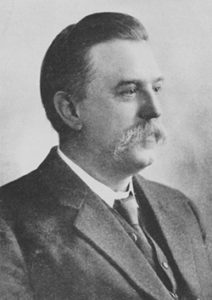 106 years ago today there appeared a 3 page report by T.K. Dalziel in the British Medical Journal (vol. 2, no. 2756, pp 1068-1070, October 25, 1913) that is widely regarded as the first report of the chronic inflammatory intestinal condition that is now called Crohn’s disease; named for the first of three authors of the 1932 report describing the pathology and clinical presentation of this regional ileitis. Dalziel describes several cases, three of which have pathology reports.
Noteworthy is that Dalziel mentions the gross and microscopic similarities of the intestines he removed from patients with those from cases of Johne’s disease in cattle. He mentions the 1895 work of Johne (misspelled as Henny) and Frothingham and cites the early work on the Johne’s disease pathology by McFadyen. The puzzling aspect is that while the pathology of the human and animal diseases is strikingly similar, acid-fact bacteria (MAP) can be seen in the animal tissues but not in the human tissues: a puzzle that remains today and is the essential feature leading some experts to view the human and animal forms of this chronic enteritis as having different causes.
Comment: It is often useful to read the original published reports to avoid perpetuating misunderstandings. However, it is often hard to obtain older literature. It is for this reason that the Johne’s Information Center highlights provides access this seminal report. This is made possible by JSTOR (short for journal storage) and we are grateful to this organization for making the article accessible.
There were 6.8 million cases of inflammatory bowel disease (IBD) globally in 2017. At the national level, the USA had the highest age-standardized prevalence rate (464 per 100,000 population), followed by the UK (450 per 100,000). This equates to roughly 1 out of every 220 people having IBD. The total years lived with disability (YLDs) attributed to IBD almost doubled over the study period, from 0.56 million in 1990 to 1.02 million in 2017. These data come from a study funded by Bill & Melinda Gates Foundation published online October 21 in Lancet Gastroenterology Hepatology. This excellent Open Access article is titled: “The global, regional, and national burden of inflammatory bowel disease in 195 countries and territories, 1990–2017: a systematic analysis for the Global Burden of Disease Study 2017”.
IBD in humans and Johne's disease (JD) in animals have both increased in prevalence globally over the past 100 years. The prevalence of IBD and JD by country are also strikingly similar. Read more about the zoonotic potential of MAP and the association of MAP and IBD here.
TESTING HUMANS FOR MAP
2019-10-20 15:29:06 Human Para’s inaugural MAP testing study is nearing completion. A total of 201 participants donated a blood sample at locations in Orlando, Philadelphia and New York City between May and September 2018. The final samples were drawn on September 10, 2018.
Since this was a blinded study comprised of both IBD patients and control subjects, each sample was assigned a number and sent to 6 participating researchers. The laboratories of Dr. Saleh Naser, Dr. Tim Bull and Dr. Irene Grant received buffy coats (the white blood cell layer) which were extracted at the Temple University laboratory. They tested all positive culture samples for two PCR markers unique to MAP: IS900 and f57. More detail about the testing methodologies can be found at the Human Para Foundation website.
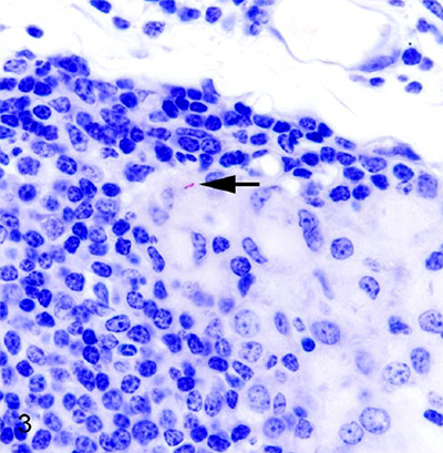
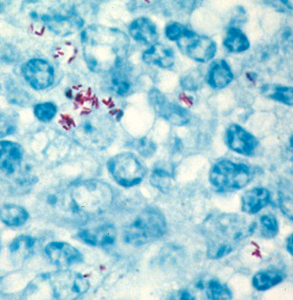
Comment: Zoonotic pathogens typically have their “preferred” normal host. When they infected a different, abnormal host, they often change their behavior. When MAP finds itself inside a host that is not the normal one, i.e. not a ruminant animal, it may behave in unpredictable ways. Some experts suggest that it stops making its typical thick waxy cell wall rendering it difficult to stain using normal acid-fast stains for visualizing mycobacteria and no longer looks like a rod-shaped bacterial cell. This may explain why pathologists cannot see typical acid-fast stained (red) rod shaped bacteria as shown below in a tissue section from a cow.
Diagnostic tests for MAP in humans therefore may necessarily differ in design from those tests used in animals. Hopefully, among the 6 different testing methods being evaluated at least one emerges that can effectively identify MAP infections in humans. An accurate diagnostic test for MAP in humans combined with the effective anti-MAP therapy being pioneered by RedHill Biopharma could revolutionize diagnosis and treatment of Crohn’s disease and other disease of humans linked to MAP.
LANDMARK PAPER DEFINING CROHN’S DISEASE (1932)
2019-10-18 13:29:51 Eighty-seven years ago, Burrill B. Crohn, Leon Ginzburg, and Gordon D. Oppenheimer published a paper titled Regional Ileitis – A Pathologic and Clinical Entity in the Journal of the American Medical Association (vol. 99, no. 16, pp 1323-1329, October 15, 1932). Honoring the importance of this report, the article was later reprinted as a Landmark Article in The Mount Sinai Journal of Medicine (vol 67, no. 3, pp 263-268, May 2006). We provide the original JAMA article here for users interested in reading this influential publication in its original form. Note: the reprinted version in the Mount Sinai Journal of Medicine has better print quality.
Comment: I appreciate history more as I grow older. Also, it is important to read original published reports to avoid misquoting or perpetuating misunderstandings. Interesting note: As described in Wikipedia, Crohn always preferred the medically descriptive terms "regional ileitis" and "regional enteritis" to "Crohn's disease", but he was not able to prevent the appropriation of his name for the disease.
Without providing much detail, B.B. Crohn’s article mentions efforts to determine if Mycobacterium tuberculosis was involved in the regional ileitis cases he described including culture for M. tuberculosis, inoculation of lymph node homogenates from five patients into guinea pigs, rabbits, and chickens, and acid-fast staining of tissue sections. He concludes that M. tuberculosis was not a cause of these cases of regional ileitis. However, he never mentions the 1913 report by Dalziel or makes any mention of Mycobacterium paratuberculosis or the similarities of regional ileitis in humans to that of cattle, as described by H.A. Johne in 1895. Clearly, Dr. Crohn recognized how the pathology in his afflicted patients resembled that caused by a mycobacterial infection. How might history be different had Dr. Crohn considered the possibility M. a. paratuberculosis was the cause?
Johne's disease is a regional ileitis affecting ruminants.
Dr. Robert Greenstein compared Crohn's disease and Johne's disease in his "personal view" article published in Lancet Infectious Diseases in 2003 (Lancet Infect Dis 2003; 3:507-14).
If you are interested in the history of Johne's disease, check out our timeline of major events.
The picture of B.B. Crohn is credited to Wikipedia.
« Previous 1 … 11 12 13 14 15 … 18 Next »