
MAP-INFECTED CARNIVORES IN PORTUGAL
2020-01-26 18:11:07 Today's news: Another report of probable MAP “spillover” from domestic animals to wildlife. Monica Cunha from the National Institute for Agrarian and Veterinary Research, Oeiras, Portugal and colleagues report detection of MAP by PCR in 10% red fox (Vulpes vulpes), 6% Egyptian mongoose (Herpestes ichneumon) as well as stone marten (Martes foina) and common genet (Genetta genetta) – a first time report of MAP in genet. This Open Access publication appeared January 21 in Nature.com, Scientific Reports. [14 pages with 88 references]
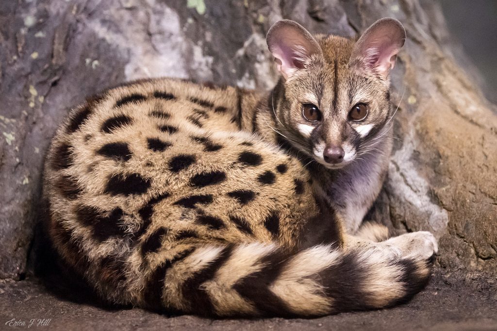
Common genet photo from Wikipedia.
Abstract
Mycobacterium avium subsp. paratuberculosis (MAP) is the etiological agent of Johne’s disease or paratuberculosis, a chronic infection affecting domestic ruminants worldwide. Despite sporadic reports of MAP occurrence in non-ruminants, information on the risk factors predisposing for infection is still scarce and evidence of transmission paths linking the livestock-wildlife-environment interfaces also remains lacking. In this study, we predicted that environmental, host-related, land use and human driven disturbance factors would modulate carnivore exposure to MAP. To test these hypotheses, we performed a retrospective survey, based on microbiological and molecular methods, in mainland Portugal including five sympatric species from the Herpestidae, Canidae, Viverridae, and Mustelidae families (n = 202) and examined 16 variables as putative predictors of MAP occurrence. Molecular evidence of MAP using IS900 as proxy was demonstrated in 7.43% (95%CI: 4.55–11.9) of surveyed carnivores, the highest proportions being registered for red fox (Vulpes vulpes) (10%; 95%CI: 4.0–23) and Egyptian mongoose (Herpestes ichneumon) (6.0%; 95%CI: 3.2–11). We demonstrate that important species of the Mediterranean carnivore guild, such as stone marten (Martes foina) and common genet (Genetta genetta), may also be exposed to MAP, being this the first time that occurrence in genet is reported. The high proportion of DNA-positive specimens, concurrent with the apparent lack of gastro-enteric lesions and molecular confirmation of IS900 in feces, argue for the presence of subclinical carriers that occasionally shed bacteria, potentially aiding as source of infection to susceptible species and possibly contributing for environmental contamination. Achievement of MAP isolation would prove beyond any doubt that MAP is present in this wildlife population. Ecological modelling results suggested that the probability of MAP infection using IS900 as proxy in mongoose is positively associated with higher altitude and temperature stability, as well as with lower annual rainfall. Density of livestock farms was found not to be a significant predictor, which may indicate that the livestock-wildlife interface is probably not important as an infection route for mongoose.
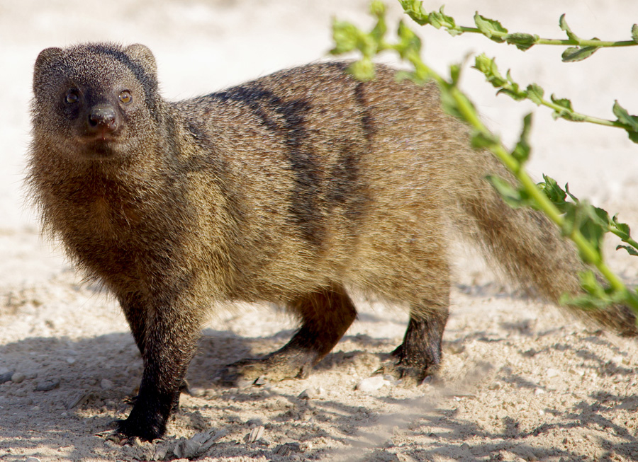
Egyptian mongoose photo from Wikipedia.
Concluding remarks excerpt: Suspicion for the ecological spread of MAP reinforces the need for increased surveillance and action. Quantitative information on the prevalence and concentration of MAP in putative contaminated sources of exposure is required.
Comment: This report adds to the growing body of literature about MAP spread to wildlife. It is critical to establish whether the transmission of MAP is a “two-way street”, with MAP going back and forth between domestic animals and wildlife, or instead that wildlife are a “dead-end host”. i.e. transmission only from domestic animals to wildlife ("one-way street"). The answers will likely depend on the type of wildlife, i.e. wild ruminants probably develop permissive infections shedding MAP in ways that can then infect domestic animals, while wild carnivores may be dead-end hosts. Read more about non-ruminants that have been found to be MAP-infected on this website page.
DAIRY GOATS THREATENED BY JOHNE’S
2020-01-20 19:41:18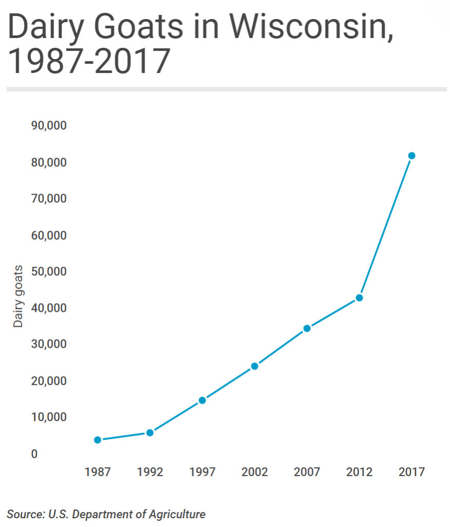
As reported in the Wisconsin State Farmer, September 16, 2019:
Wisconsin's self-proclaimed moniker as "America's Dairyland" is taking on fresh meaning in the 21st century thanks to a growing market for milk from an animal that bleats rather than moos. While the state's traditional dairy cattle industry continues to hemorrhage producers at a record pace, Wisconsin's dairy goat industry is in the midst of a long-term, and accelerating, growth spurt. Indeed, in 2019 Wisconsin can reasonably claim to be America's dairy (goat) land.
Data from the United States Department of Agriculture, which counts livestock across the United States every 5 years, show just how much Wisconsin dominates the nation's dairy goat industry. In 2017, the most recent year the USDA surveyed producers, the size of Wisconsin's dairy goat herd easily topped the nation at more than 83,000-head. California came in a distant second, with some 43,000 dairy goats, while Iowa, Texas and Missouri rounded out the top five.
It's not only the sheer size of Wisconsin's dairy goat herd that stands out: The state also leads the nation in the value of sales from dairy goat operations and is the epicenter of national growth in goat dairy.
Read the rest of this excellent article that contains many useful links here.
Comment:
Rapidly expanding animal industries commonly overlook biosecurity risks feeling compelled to buy animals regardless of health status. Many of these new dairy goat operations have 5,000 to 10,000 goats. Operations only can attain this size by purchase of goats from many different herds. Purchasing animals is the #1 way by which herds, whether of cattle, sheep or goats, become infected with MAP. This then leads to long-term problems of animal health, animal welfare, animal productivity and animal longevity which all translate to a negative economic impact due to Johne’s disease.
Johne’s disease is common among all goat breeds.
- France found that 55.2% of dairy goat herds had one or more ELISA-positive goats.
- In Cyprus 50% of dairy goat herds were seropositive.
- In Ontario in Canada 83% (estimated true prevalence) of dairy goat herds were MAP-infected.
- In Spain 87.5% of dairy goat herds were seropositive.
- I found no such surveys in U.S. dairy goats but a study of Boer goats in Missouri reported that an estimated 54.7% of Boer goat herds are seropositive for Johne’s disease.
Many owners fail to realize their goats are MAP-infected and therefore do not implement control measures with the result that the infection continues spreading. In this article in the Wisconsin State Farmer, I also observed that links provided in this article to goat associations and experts never once mention biosecurity concerns or the threat of Johne’s disease. So unfortunate!
Goat owners and veterinarians must be much more aware of Johne’s disease and implement biosecurity protocols and on-farm control measures to mitigate the damage caused by Johne’s disease. When MAP becomes recognized as a food-borne zoonotic pathogen, these large goat herds with face a daunting task to control or eradicate this infection.
The age-old wisdom that “prevention pays” should be heeded.
MAP THREATENS ENDANGERED DEER
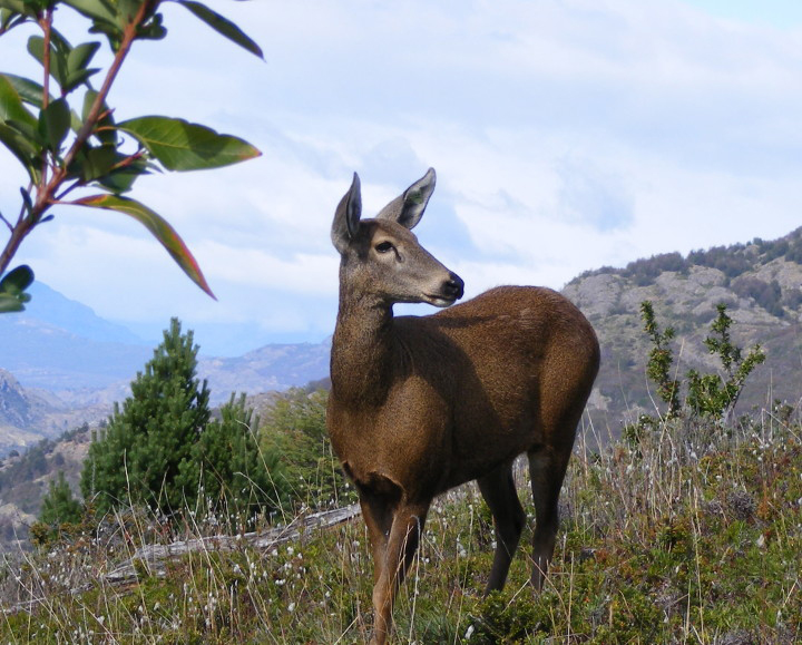
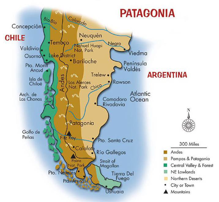
2020-01-11 19:57:31A rare and beautiful sight, the Southern Patagonian huemul deer (Hippocamelus bisulcus) is an icon of Patagonia Park and of Chile. A native species, the huemul is Chile’s national animal, found on the country’s coat of arms, alongside the Andean condor. Despite its high level of recognition, the huemul is classified as endangered by the IUCN, with a global population of less than 1,500 individuals. With 10% of these remaining deer residing within Patagonia Park’s boundaries, Conservacion Patagonica has made huemul deer recovery the cornerstone of its wildlife program.
Paulo Corti and colleagues from Universidad Austral de Chile, Valdivia, Chile describe recovery of MAP from 3 of three fecal samples from huemul deer in Patagonia in The Australian Journal of Veterinary Science (52:33-35, 2020).
Abstract
In a huemul (Hippocamelus bisulcus) population sympatric with cattle, we found evidence of Mycobacterium avium subsp. paratuberculosis (MAP) infection. Three huemul faecal pellet samples and two cow pats were collected and cultured for MAP presence. DNA was then extracted for PCR analysis of all signal-positive cultures. To assess whether MAP isolates obtained from huemul faeces were associated with typical MAP isolated from livestock, positive confirmed culture samples were sub-typed using a combination of five Mycobacterial Interspersed Repetitive Unit-Variable Number Tandem Repeat Analysis and one Short Sequence Repeat analysis markers. All faecal samples from both species were MAP positive. One huemul presented a different bacteria profile genotype not described before, suggesting that huemul and cattle in Patagonia could carry a unique MAP strain.
Comment: The primary reservoir for MAP is in domesticated ruminants such as dairy cattle, beef cattle, sheep and goats. However, MAP infections in these livestock readily spread to wild ruminants and this first-time report of finding MAP in huemel deer illustrates the concept of infection “spillover”.
 Spillover is the title of an excellent book by David Quamenn which focuses on the spillover of infectious diseases from animals to humans, so called zoonotic infections. MAP infections clearly spillover to wild ruminants, and non-ruminants, including humans. This website provides more details about MAP infections of dairy cattle, beef cattle, goats, sheep, deer & elk, bison, water buffalo, wild ruminants, zoo ruminants, and non-ruminants. A separate page is devoted to the zoonotic potential of MAP.
MAP is a promiscuous, insidious, zoonotic, foodborne pathogen that threatens the economic viability of livestock producers, the health and well-being of wildlife, zoological collections of wild ruminants (many of which are endangered), and humans. It deserves far more research funding and control program investment than it currently receives.
SHEEP AND SAMPLE POOLING
2020-01-05 17:33:52Research Report
Mathevon and colleagues from Ecole Nationale Vétérinaire de Toulouse, Toulouse Cedex, France reported on optimal sheep sample pooling strategies for serum samples (for ELISA testing) or fecal samples (for qPCR testing). Their report appeared December 26, 2019 in PLoS ONE and is OPEN ACCESS. [18 pages with 47 references]
Abstract
The aim of our study was to evaluate the flock sensitivity and specificity of fecal qPCR and serum ELISA using pooled samples for screening paratuberculosis in French sheep.
Using individual feces with low or high qPCR Ct values from ewes sampled in 14 infected flocks, a total of 555 pools of size 5, 10 and 20 were created by diluting individual materials in negative feces and analysed using a commercial IS900 qPCR kit. The relative performances of pooled serum ELISA analysis were evaluated based on the analysis of 181 different pools of size 5 and 10, composed of individual serum samples of various individual S/P values. Results showed that for pools of size 5, 10 or 20, individual fecal samples with low Ct values were invariably detected. Conversely fecal samples with high Ct values were associated with a lower detection rate in both pools of size 5 (87.0% to 90.0%), 10 (63.0% to 70.7%) and 20 (46.7% to 60.0%). After lowering the decision threshold to 25% and 15% for serum pools of size 5 and 10 respectively, the pooled serum ELISA relative sensitivity ranged between 62.2% and 100.0% depending on the composition of the pools.
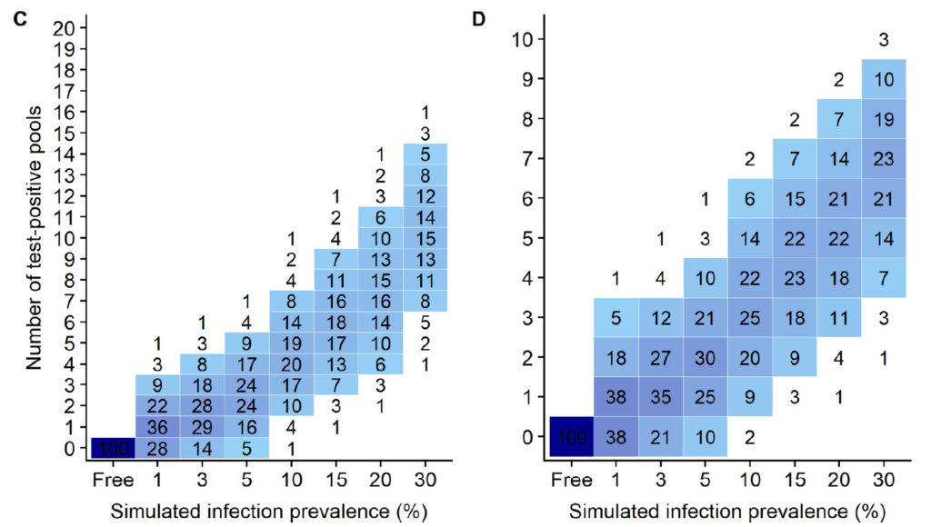
Finally, a simulation study was carried out to evaluate the performances of 16 screening strategies at flock level, with varying pool size (5 to 20) and number (5 to 60). The use of pooled serum ELISA led to very false positive detection rate ranging between 37.6% and 91.8% in paratuberculosis free flocks and prevents its further use in that context. For infection prevalence ≤ 5%, the flock sensitivity based on pooled fecal qPCR ranged between 39.0% (5 pools of size 10) and 99.9% (300 sampled individuals, with pools of size 5,10 or20), and was always above 93% when the infection prevalence was greater or equal to 15%. We conclude that pooled-fecal qPCR but not pooled-serum ELISA could be a useful tool to detect sheep flocks infected with paratuberculosis.
Comment: The authors focused their discussion on diagnostic tests useful for screening of sheep flocks for ovine Johne’s disease (OJD) where the primary goal is to detect MAP-infected flocks, i.e. maximize “flock sensitivity” and “flock specificity”, at the least cost. This should not be confused with the use of fecal pooling strategies for qPCR testing in known infected flocks where the goal is to identify individuals in the flock that should be culled or isolated.
In MAP-infected flocks there are two equally important goals: 1) to limit the cost of testing the whole flock, and 2) to limit the cost of testing the individual samples, i.e. to have the fewest number of qPCR-positive pools. This second goal is important because each of the fecal samples comprising a qPCR-positive pool must be individually tested by qPCR in order to identify each MAP-infected sheep adding significant costs, e.g. roughly $30 x 5 = $150 per PCR-positive pool (based on WVDL testing rates).
Using the simulation data from this publication (shown above), for flocks with a MAP infection prevalence of 10%, the simulation data (Fig 4 C & D in the publication as shown above) indicate that using 20 pools of 5 fecals/pool, 93% of flocks would have 7 or fewer positive pools which would then cost an additional $1,050 to test each fecal individually by qPCR (7 pools x 5 fecals/pool x $30/qPCR = $1,050). By contrast, if these same flocks used 10 pools of 10 fecals/pool, then 92% of flocks would have 5 or fewer qPCR-positive pools resulting in a cost of $1,500 to individually test the fecal samples making up those pools (5 pools x 10 fecals/pool x $30 = $1,500).
The cost of testing by pooled PCR at the WVDL is $34/pool so would cost $340 for 10 pools of 10 fecals/pool or $680 for 20 pools of 5 fecals/pool. Thus, while testing 20 pools (5 fecals/pool) would initially cost $340 more, the cost of testing the individual fecals comprising each qPCR-positive pool does not offset the cost of testing fewer pools in this example.
These findings are based on a simulation of random pooling of fecal samples described in the publication. As mentioned in a previous news posting, strategic pooling by animal age can help limit the number of test-positive pools Kalis (2000). This is because animals of similar age experienced similar risks of becoming MAP-infected when young. Thus, age-based pooling (strategic) results in more MAP-infected animals likely to be in the same pool and conversely the fewest number of positive pools for the flock or herd as a whole.
You can read more about a clustering of MAP-infections by age and season in the publication by Zare et al. titled: Evidence of birth seasonality and clustering of Mycobacterium avium subspecies paratuberculosis infection in US dairy herds. Preventive Veterinary Medicine 112: 276– 284, 2013. However, the clustering effect may not be as pronounced for sheep which do not have lambs thought the year.
In closing, this discussion highlights the importance of utility analysis when judging the merits of diagnostic tests for MAP. For more on this topic see Garder et al. Consensus-based reporting standards for diagnostic test accuracy studies for paratuberculosis in ruminants. Preventive Veterinary Medicine 101: 18-34, 2011.
MAP IN BRAZILIAN CHEESE
2019-12-30 15:39:49Research Report
Brazilian researchers report a small survey of a popular cheese eaten in Brazil for MAP. Their findings were reported in Arquivo Brasileiro de Medicina Veterinária e Zootecmia (vol.71 no.6 Belo Horizonte Nov./Dec. 2019 Epub Dec 13, 2019).
Abstract
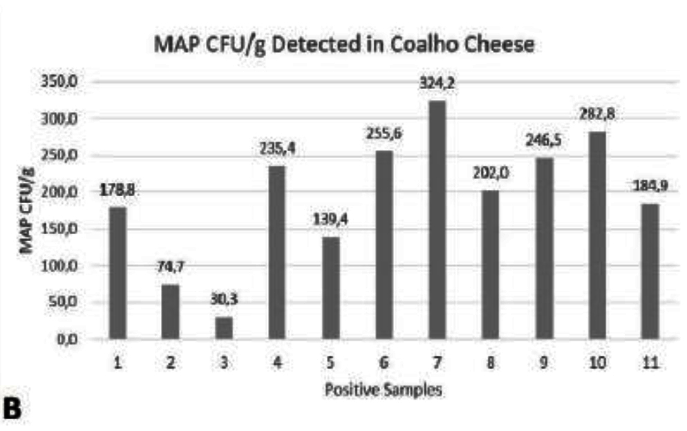
Paratuberculosis is a chronic and incurable disease that affects ruminants and other domestic animals. It is caused by Mycobacterium avium subsp. paratuberculosis (MAP) that may also be involved in some human diseases such as Crohn's disease, type 1 diabetes, sarcoidosis, multiple sclerosis, and Hashimoto's thyroiditis. The objective of this study was to investigate the occurrence of MAP DNA in samples of artisanal coalho cheese purchased in the State of Pernambuco. Forty samples of coalho cheese submitted to the Real Time Polymerase Chain Reaction (qPCR) technique were analyzed for the detection of the MAP region IS900. 11 (27.5%) were positive with a mean of 195.9 MAP colony forming unit (CFU) per gram of each sample, with a minimum of 30.3 CFU/g and a maximum of 324.2 CFU/g. Thus, this type of cheese that is one of the most consumed in this region of Brazil constitutes a source of human exposure to MAP. Further research in this area should be performed to evaluate the viability of the bacteria in this cheese type.
Comment: It should be noted that MAP numbers (CFU) were actually based 10-fold serially diluted MAP DNA used to obtain the standard curve, in which it was possible to detect from 21 up to 2.12 x 10⁷ MAP cells. In my opinion a more informative titration curve (MAP CFU vs qPCR Ct values) would begin with known CFU/mL MAP suspensions that were then processed by DNA extraction followed by qPCR. Only then can an investigator truly know the net results of both DNA extraction and target IS900) amplification to estimate the CFU of MAP in the original sample. That said, most molecular biologists opt for titration of extracted DNA as the authors of this study did.
The last line of the abstract is the critical one. The really big question is: Are the detected MAP alive or dead. That said, some investigators postulate that dead MAP may function as a dietary allergen triggering inflammation in the absence of infection. MAP as a food safety threat is a critical knowledge gap urgently requiring research.
MAPPING MAP IN MINNESOTA
2019-12-23 13:04:54Research Report
KST Kanankege and colleagues from the Department of Population Medicine, College of Veterinary Medicine, University of Minnesota reported on use of milk ELISA testing data from a voluntary testing program through DHI testing laboratories as a passive surveillance tool for Johne’s disease in Minnesota. Their report appears in the journal BMC Veterinary Research 15:429, 2019. [Open Access]
Abstract
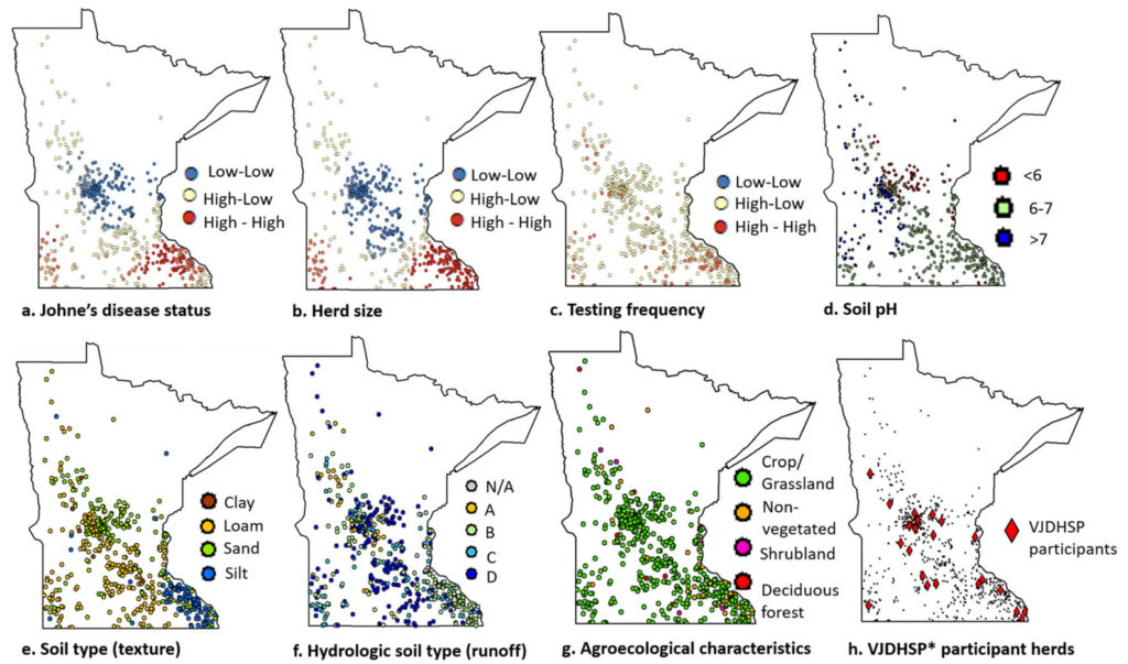
Background: One of the key steps in the management of chronic diseases in animals including Johne’s disease (JD), caused by Mycobacterium avium subsp. paratuberculosis (MAP), is the ability to track disease incidence over space and time. JD surveillance in the U.S. dairy cattle is challenging due to lack of regulatory requirements, imperfect diagnostic tests, and associated expenses, including time and labor. An alternative approach is to use voluntary testing programs. Here, data from a voluntary JD testing program, conducted by the Minnesota Dairy Herd Improvement Association, were used to: a) explore whether such a program provides representative information on JD-prevalence in Minnesota dairy herds, b) estimate JD distribution, and, c) identify herd and environmental factors associated with finding JD-positive cows. Milk samples (n = 70,809) collected from 54,652 unique cows from 600 Minnesota dairy herds between November 2014 and April 2017 were tested using a MAP antibody ELISA. Participant representativeness was assessed by comparing the number of JD-tested herds with the number of herds required to estimate the true disease prevalence per county based on official statistics from the National Agricultural Statistical Services. Multivariable logistic regression models, with and without spatial dependence between observations, were then used to investigate the association between herd status to JD (positive/negative), as indicated by milk ELISA results, and available covariates at the herd level.
Results: Within the study population, at least one test-positive cow was found in 414 of 600 (69%) herds. Results indicated that large herds that test frequently and herds located in loamy or silt soils are more likely to have at least one MAP test-positive cow. After adjusting for herd size, testing frequency, and soil type, there was no spatial dependence in JD risk between neighboring dairies within 5 to 20 km. Furthermore, the importance of collecting data on herd management, feed, and biosecurity for insightful interpretations was recognized. The study suggested that, although limited, the voluntary testing database may support monitoring JD status.
Conclusions: Results presented here help elucidate the spatial characteristics of JD in Minnesota and the study may ultimately contribute to the design and implementation of surveillance programs for the disease.
Comments: This excellent article demonstrates the power of utilizing diagnostic testing data for Johne’s disease to explore relationships and environmental factors affecting the prevalence of Johne’s disease. This is data that otherwise would provide no benefit to the dairy industry and simply sit idly on computer hard drives. Kudos to the Minnesota Dairy Herd Improvement Association for their support in data collection! Imagine if all DHI labs and all state veterinary diagnostic labs could/would share data in this same way to provide a comprehensive understanding of the spatial distribution and risk factors affecting Johne’s prevalence in the U.S.: a relatively small investment with potentially big returns.
OPTIMAL FECAL POOL SIZE
2019-12-15 18:44:50Research Article
Australian veterinary scientists report studies to define the optimal number of fecal samples from beef cattle that can be pooled for MAP detection by fecal PCR. Their findings were published in PLoS ONE and the article is Open Access [18 pages with 42 references]. This was mentioned in last weeks news posting but I feel it deserves more explanation.
Abstract
Bovine Johne’s disease (JD) is a chronic debilitating disease affecting cattle breeds worldwide. Pooled faecal samples are routinely tested by culture to detect Mycobacterium avium subsp. paratuberculosis (Mptb) [also called MAP] infection. More recently, a direct high throughput molecular test has been introduced in Australia for the detection of Mptb in faeces to circumvent the long culture times, however, the optimal pool size for beef cattle faeces is not known. This study aimed to determine the optimal pool size to achieve the highest test sensitivity and specificity for beef cattle. Individual archived faecal samples with low, medium and high quantities of Mptb (n = 30) were pooled with faecal samples from confirmed JD negative animals to create pool sizes of 5, 10, 15 and 20, to assess the diagnostic sensitivity relative to individual faecal qPCR. Samples from JD-free cattle (n = 10) were similarly evaluated for diagnostic specificity. Overall, 160 pools were created, with Mptb DNA extracted using magnetic bead isolation method prior to Mptb-specific IS900 quantitative PCR (qPCR). The pool size of 10 yielded the highest sensitivity 73% (95% CI: 54–88%), regardless of the quantity of Mptb DNA present in the faeces. There was no significant differences between the four different pool sizes for positive pool detection, however, there was statistical significance between low, medium and high quantities of Mptb. Diagnostic specificity was determined to be 100%. The increase in pool size greater than 10 increased the chances of PCR inhibition, which was successfully relieved with the process of DNA dilution. The results of this study demonstrate that the pool size of 10 performed optimally in the direct faecal qPCR. The results from this study can be applied in future simulation modelling studies to provide suggestions on the cost-effective testing for JD in beef cattle.
Comment: Dr. Kees Kalis (2000) also showed that pooling of 5 fecal samples was effective when using culture as the diagnostic test. Kalis also noted that “strategic pooling” can further enhance the procedure. Strategic polling refers to grouping animals of the same or similar ages into the same pools.
A study on fecal sample pooling by Wells et. al. (2002) using artificially constructed pools of feces from dairy cattle showed that 5 samples per pool was only slightly better than 10 samples per pool, when using culture of MAP on solid media as the diagnostic test. The standard protocol for sample pooling in the U.S. has remained at 5 per pool ever since.
The Australian study used a much larger number of samples, they originated from naturally infected beef cattle, and the diagnostic assay was the high throughput PCR (Plain, 2014). The ability to pool 10 samples instead of 5 obviously decreases the laboratory costs for the producer by half with apparently little or no loss in diagnostic accuracy. If widely adopted, this would encourage more testing and verification of cattle herds that are not MAP-infected which then would be preferred sources of replacement cattle. These cattle could sell at premium prices thus allowing the herd owner to recoup the costs of attaining a test-negative status.
Australia has been using fecal sample pooling for sheep since 2000 finding that fecal pellets from 50 sheep can be combined into a single pooled test without significant loss in MAP infection detection at the flock level (Whittington, 2000).
Due to lack of funding, few cattle herds currently participate in the US. JD program (formally called the Voluntary Johne’s Disease Herd Status Program (VJDHSP). Thus, for herd owners simply wanting the least cost JD surveillance program, without concern for formal USDA recognition of the herd JD status, then it makes sense to use the 10 fecals/pool testing scheme.
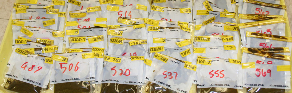
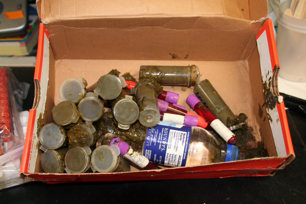
Lastly, samples should be carefully collected, packaged and shipped refrigerated to the lab. The containers should seal tightly to prevent leakage. Below is an example of how NOT to send fecal samples. These snap-top containers were filled too full and not refrigerated causing gas to be produced popping the tops off the containers making for a poopy mess and cross-contamination of samples rendering them useless. The photo above shows well-collected and labeled bovine fecal samples in Whirlpack bags.
TESTING BEEF CATTLE HERDS
2019-12-12 14:51:33John Campbell published an article in the December 6 issue of The Western Producer titled: Testing beef herds for Johne’s disease can be difficult. Interestingly, he begins his article stating: "Every year, I seem to write another article about Johne’s disease. It is one of the most common topics of phone calls and questions that I receive from veterinarians and producers."
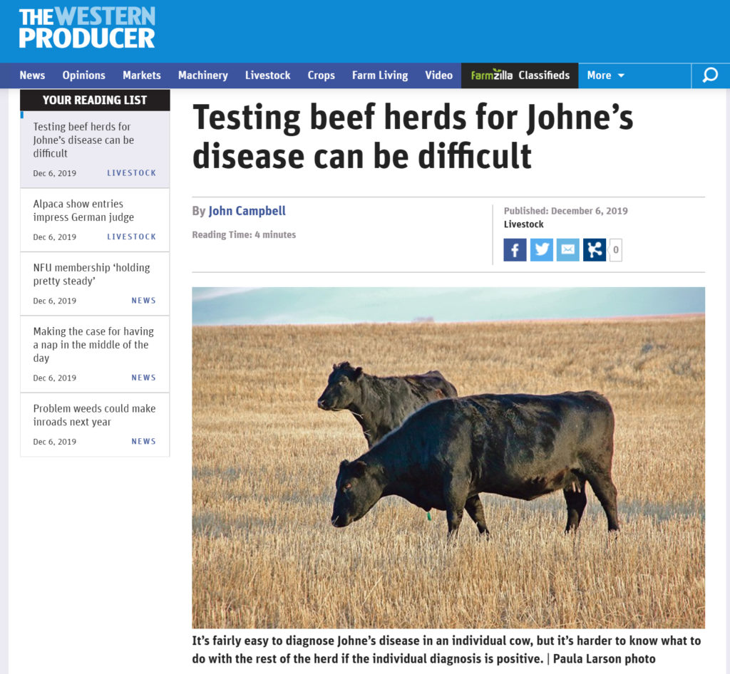
It’s very helpful that this article written for beef cattle producers calls attention to Johne’s disease. However, it is more pessimistic about diagnostic testing than necessary.
This news posting offers an alternative perspective.
Mr. Campbell describes a hypothetical 1,000 cow herd with a 10% true MAP infection rate. He analyzes what would occur using a blood test (ELISA) with a sensitivity and specificity of 30% and 99%, respectively. He explains that on a single herd test there will be too many false-negative test (7 of the 10 MAP-infected cows would be missed). In his example, he claims there also would be too many false-positive test results (9 cows that are truly not MAP-infected would test positive) resulting in too many mistakenly culled cows. There are several problems with this analysis.
First, the beef cattle herds that should be testing are those sell breeding stock, often called seedstock producers. For commercial cow-calf producers a such a testing program is good for animal health and welfare but hard to justify economically. For seedstock producers, eradicating MAP infections or proving herds are not MAP-infected must be a business objective to avoid spreading MAP infections to other herds.
Second, beef seedstock herds should not be using a blood test (ELISA). Rather they should use a test with higher sensitivity and specificity namely the direct fecal PCR assay. The high throughput PCR assay used in Australia has a reported sensitivity and specificity of 60.4% and 99.6%, respectively (Plain, 2014).
Also, ELISAs are screening tests and most experts recommend doing a confirmatory test (culture or PCR) on all ELISA-positive animals before making a decision to cull the cow. Especially if the cow is valuable.
Had Mr. Campbell used fecal PCR as the test in his example, only 4 of the 10 MAP-infected cows would be missed on a single whole herd test and these so called false-negative cows would be animals that were shedding few or no MAP in their feces, meaning they are not infectious. These animals would likely be detected in the next annual herd test. The false-positive rate would be 0.4% (1.00 minus specificity) or 3 - 4 cows (890 non-infected cows x 0.4% = 3.5).
Not only is a direct fecal PCR the test of choice, but this assay is more economical because it can be done on pooled fecal samples (the lab does the pooling). Until recently, the standard pooling method mixed fecal samples from 5 cows into a single pool that was then tested by PCR. This month, Australian researchers showed that this can be increased to 10 animal per pool (Ly, 2019).
At the Wisconsin Veterinary Diagnostic Laboratory the ELISA cost is $6/sample and the pooled PCR is $34/pool, or $6.80/cow, using pools of 5, and it would be only $3.40/cow, using pools of 10. Some additional costs would be incurred to run a confirmatory test on ELISA-positive cows and to test individual cows in the PCR-positive fecal pools.
____________
In summary, pooled fecal PCR is the test of choice for beef cattle seedstock herds. This assay provides twice the diagnostic sensitivity (rate of detecting MAP-infected cows) and higher diagnostic specificity (fewest false-positives) at a cost that is comparable to or lower than that of the ELISA blood test. Additionally, the fecal PCR provides numerical results that indicate which cows are shedding the most MAP, i.e. those that are most infectious and should be culled first. This is also true for breeders of sheep, goats, dairy cows and other ruminants.
ENVIRONMENTAL TESTING FOR MAP ON LARGE DAIRIES
2019-12-08 18:27:01Research Report
Chamchoy T, Williams DR, Adaska JM, Anderson RJ, Aly SS. 2019. Environmental sampling to assess the bioburden of Mycobacterium avium subspecies paratuberculosis in drylot pens on California dairies. PeerJ 7:e8081 https://doi.org/10.7717/peerj.8081
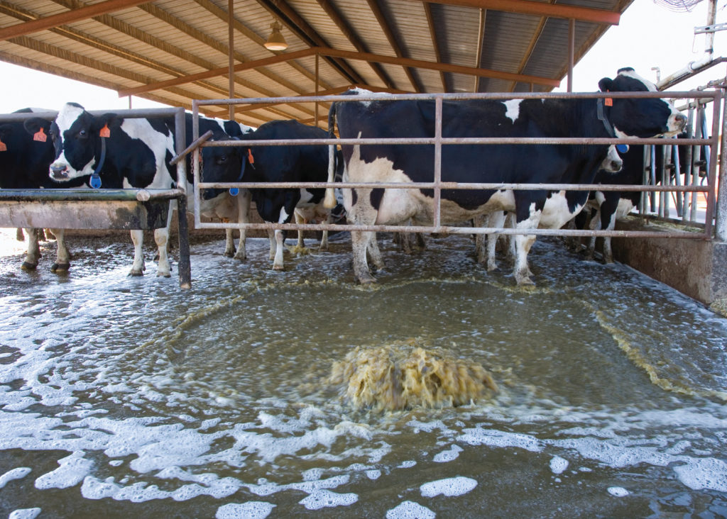
Abstract
Mycobacterium avium subspecies paratuberculosis (MAP) is a bacterium that can cause substantial economic losses in infected dairy herds due to reduced milk production and increased cow-replacement costs. In order to control MAP in dairies with drylot pens, a standardized environmental sampling protocol to quantify MAP in fecal slurry was developed based on an existing protocol for freestall pens. Specifically, following a 24 h hold of the flush, a grab sample of approximately 10 ml of fecal slurry was collected every 1 m along the flush lane of the drylot pens, avoiding individual cow fecal pats. To determine the reliability and repeatability of the new environmental sampling protocol for estimation of MAP bioburden at the pen level, two collectors simultaneously collected fecal slurry samples every day for 3 days from six drylot cow pens on two Central California dairies. During the study period no cow movement between pens was allowed with the exception of sick cows. The study herds had MAP seroprevalence of 5.8% and 3.2%, respectively, based on whole pen serum ELISA results. Variance components models for quantitative real-time PCR (qPCR) results showed samples collected from different pens on different dairies accounted for greater variability in MAP concentration (65%), while samples collected by different collectors had the least variability (0.1%). In contrast, variability in MAP concentration in environmental samples collected on different days had 25% variability. The intraclass correlation coefficient showed high reliability (93%) of environmental sampling simultaneously by different collectors. In contrast, the reliability of environmental sampling at different days was 65%, which was similar to the reliability for sampling by different collectors on different days. Investigators can expect high reliability when employing the new environmental sampling protocol along with qPCR testing of environmental samples from drylot pens.
Comment: This excellent article describes the critical variables to consider when designing surveillance programs to detect MAP infections in large drylot dairies of the type found in California, USA. The publication provides excellent details regarding the environmental sampling protocol for those interested. It is also Open Access.
NEW MAP DETECTION DIAGNOSTIC
2019-12-04 20:56:55Research Report – OPEN ACCESS
B. Swift from the department of Pathobiology and Population Sciences, Royal Veterinary College together with colleagues from the University of Nottingham and Moredun Research Institute report on a means of enhancing diagnostic sensitivity for MAP detection in blood samples by use of a bacteriophge to improve the efficiency of DNA extraction prior to performing PCR. This publication appears in the November issue of Microbial Biotechnology. PBD Biotech Ltd is the company commercializing the Actiphage® technology.
Summary
Here, we describe the development of a method that exploits bacteriophage D29 as a lysis agent for efficient DNA extraction from low numbers of mycobacterial cells. This method (Actiphage®) used in combination with PCR achieved rapid and sensitive (LOD ≤ 10 cell per ml) detection and identification of viable, pathogenic mycobacteria in blood samples within 6 h. We demonstrate that mycobacteriophage D29 can be used to detect a range of mycobacteria from clinical blood samples including both Mycobacterium tuberculosis complex and Mycobacterium avium subsp. paratuberculosis without the need for culture and confirms our earlier observations that a low-level bacteraemia is associated with these infections in cattle. In a study of M. bovis-infected cattle (n = 41), the sensitivity of the Actiphage® method was 95 % (95 % CI; 0.84–0.99) and specificity was 100 % (95% CI; 0.92–1). We further used Actiphage® to demonstrate viable Mycobacterium avium subsp. paratuberculosis is present in the blood of Johne’s infected cattle. This method provides a revolutionary new tool for the study of infections caused by these difficult to grow pathogens.
For more information contact:
Bob Lyons, GM/US, PBD Biotech, Pasadena, CA,
e: Bob@PBDBio.com p: 626 394 9771.
Comment: The diagnostic sensitivity of PCR assays is heavily dependent on the efficiency of methods to extract DNA from MAP with its thick, tough waxy cell wall. Using bacteriophages to lyse the MAP appears to be more efficient than chemical extraction methods and also allows detection of only live MAP cells since the phage will not bind to and then lyse dead MAP bacteria. Hopefully further studies by independent investigators will verify the enhanced diagnostic sensitivity of the Actiphage® system and the ability of this novel diagnostic assay to detect MAP in blood samples early in the course of a MAP infection.
« Previous 1 … 10 11 12 13 14 … 18 Next »