
EFFECT OF EXPOSURE AGE AND MAP DOSE
2020-08-31 16:16:46Mortier & colleagues from the Department of Production Animal Health, University of Calgary in Canada conducted a trial to evaluate the effect of calf age at the time of MAP exposure and dose of MAP ingested on the rate of infection progression. This publication appeared in 2014 but is so helpful in understating the epidemiology and pathogenesis of MAP infections in dairy calves it is being highlighted in today’s news positive. This Open Access article appears in the journal Veterinary Research.
Abstract
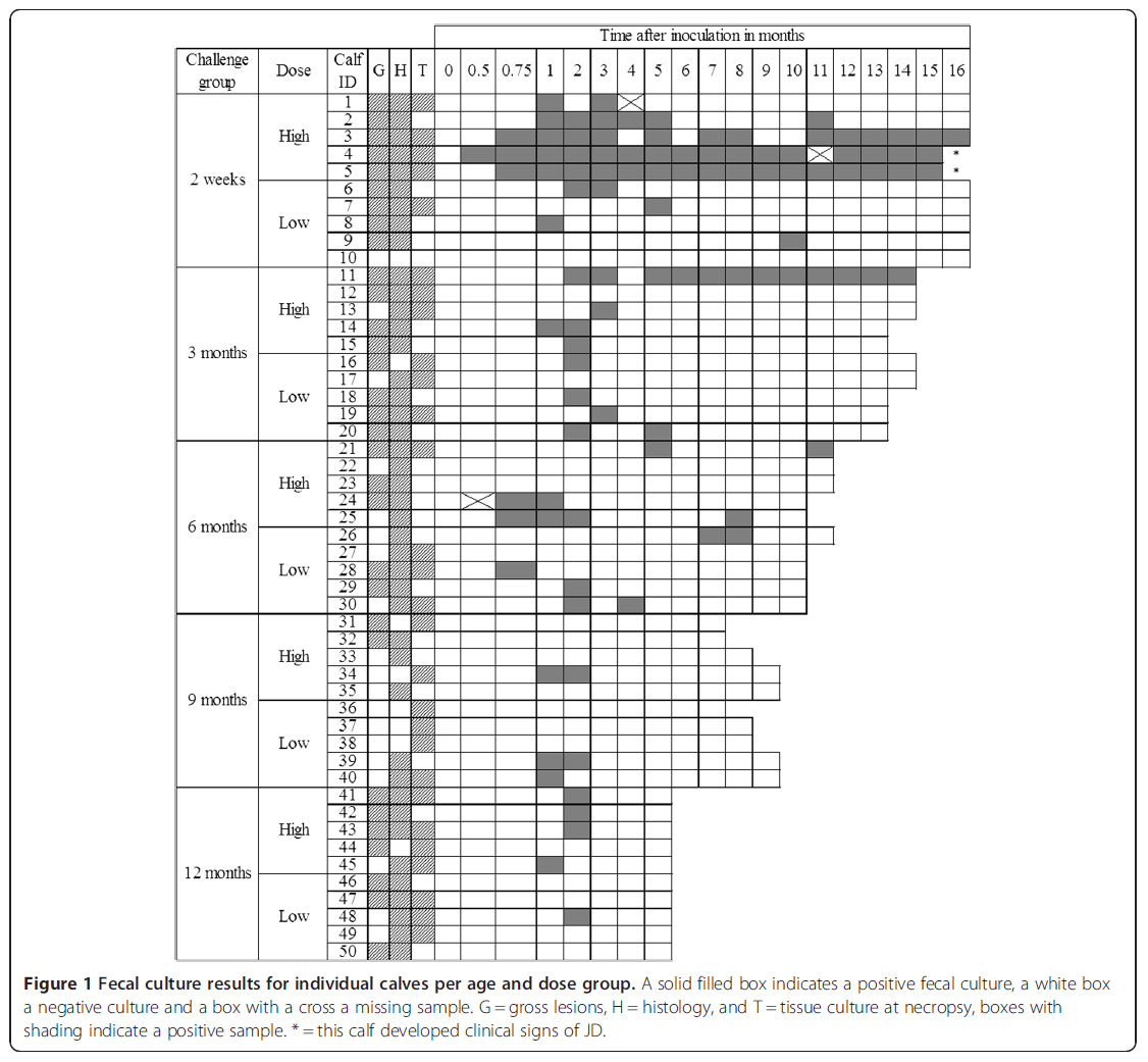
Although substantial fecal shedding is expected to start years after initial infection with Mycobacterium avium subspecies paratuberculosis (MAP), the potential for shedding by calves and therefore calf-to-calf transmission is underestimated in current Johne’s disease (JD) control programs. Shedding patterns were determined in this study in experimentally infected calves. Fifty calves were challenged at 2 weeks or at 3, 6, 9 or 12 months of age (6 calves served as a control group). In each age group, 5 calves were inoculated with a low and 5 with a high dose of MAP. Fecal culture was performed monthly until necropsy at 17 months of age. Overall, 61% of inoculated calves, representing all age and dose groups, shed MAP in their feces at least once during the follow-up period. Although most calves shed sporadically, 4 calves in the 2-week and 3-month high dose groups shed at every sampling. In general, shedding peaked 2 months after inoculation. Calves inoculated at 2 weeks or 3 months with a high dose of MAP shed more frequently than those inoculated with a low dose. Calves shedding frequently had more culture-positive tissue locations and more severe gross and histological lesions at necropsy. In conclusion, calves inoculated up to 1 year of age shed MAP in their feces shortly after inoculation. Consequently, there is potential for MAP transfer between calves (especially if they are group housed) and therefore, JD control programs should consider young calves as a source of infection.
Comment: Since calves shed MAP in feces soon after infection, it’s possible that they can transmit the infection to other calves that they are in contact with. Rather than change management practices to limit calf to calf contact, it seems more rational to invest efforts to prevent calves from becoming MAP-infected in the first place. This is done by: 1) testing the adult herd regularly, 2) culling or isolating infected cows, 3) calving test-negative cows in clean maternity pens, 4) promptly removing calves from cows, and 5) insuring colostrum fed to calves is collected only from test-negative cows and that it is collected with utmost care to avoid fecal contamination.
IRISH JD PROGRAM
2020-08-22 17:06:56Jordan et al. published a thoughtful assessment of the Irish Johne’s disease Program launched in 2018. Their Open Access article was published in the Irish Veterinary Journal August 14, 2020. This is valuable reading for countries thinking of developing comparable programs or revising their existing national program.
Abstract
The Irish dairy industry has established a reputation for the production of safe and healthy dairy products and is seeking to further expand its export market for high value dairy products. To support its reputation, stakeholders aim to control Johne’s disease. To assist decision-makers determine the most appropriate design for an Irish programme, a narrative review of the scientific literature on the epidemiology of Johne’s disease, and selected control programmes throughout the world was undertaken. Two modelling studies specifically commissioned by Animal Health Ireland to assess testing methods used to demonstrate confidence of freedom in herds and to evaluate a range of possible surveillance strategies provided additional information. The majority of control programmes tend to be voluntary, because of the unique epidemiology of Johne’s disease and limited support for traditional regulatory approaches. While acknowledging that test performance and sub-clinical seronegative shedders contributes to the spread of infection, a range of sociopolitical issues also exist that influence programme activities. The paper provides a rationale for the inclusion of a Veterinary Risk Assessment and Management Plan (VRAMP), including voluntary whole herd testing to identify infected herds and to support assurance-based trading through repeated rounds of negative testing, national surveillance for herd-level case-detection, and improved understanding of biosecurity management practices. Identification and promotion of drivers for industry and producer engagement in Ireland is likely to guide the future evolution of the Irish Johne’s Control Programme (IJCP) and further enhance its success. The provision of training, education and extension activities may encourage farmers to adopt relevant farm management practices and help them recognize that they are ultimately responsible for their herd’s health and biosecurity.
Comment: As described in the article by Jordan et al., VRAMPs are a tool to evaluate individual farm risk for the introduction and spread of MAP and educate producers on steps they can take to reduce these risks. They are typically conducted by trained private veterinary practitioners and involve an on-farm questionnaire on the management practices pertinent to MAP control. The responses to the questionnaire enable private veterinary practitioners to determine the sources of risk for the entry and spread of MAP in a herd. Farm-specific recommendations for MAP prevention and control are developed in conjunction with the farmer.
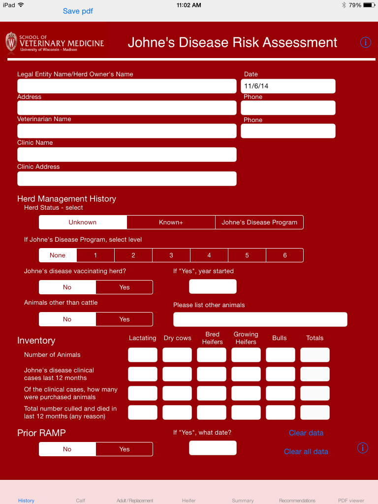
Many countries have a risk assessment and management plan (RAMP) as a component of their national program. The University of Wisconsin School of Veterinary Medicine, Food Animal Medicine Program has converted the risk assessment forms used in the U.S. national program into an App for iPads. It is one of 16 such Apps used for various health assessments in dairy herds.
DAIRY: MILK ELISAS
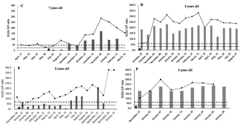
2020-08-15 16:03:46Dr. Conor McAloon from the Section of Herd Health and Animal Husbandry, School of Veterinary Medicine, University College Dublin, Ireland, together with 10 other colleagues analyzed data from dairy herds in the national Johne's Disease Control Programme (JDCP) in Ireland from January 2014 to December 2015 inclusive, consisting of 42,657 milk recordings from 18,569 cows across 187 dairy herds. Their publication in the Journal of Dairy Science appeared online July 31, 2020.
Abstract
Antibody-detecting tests for Mycobacterium avium ssp. paratuberculosis (MAP) have low sensitivity and imperfect specificity for detection of infection. Sensitivity increases as the disease progresses. Aside from infection status and stage of disease, several factors affect test performance. These factors have not yet been studied in dairy cows producing lower volumes of milk with higher solids concentration, such as those managed in low-input, pasture-based production systems. Furthermore, the effect of correcting for these associations on individual and herd test status is also unknown. The first objective of this study was to examine the relationship between MAP antibody response in milk and milk yield, somatic cell count (SCC), fat and protein contents, and stage of lactation in dairy cows enrolled in the national Johne's Disease Control Programme (JDCP) in Ireland. The second objective was to examine the effect of correcting the antibody response for these associations on the test status of individual cows and herds, given that individual tests are often used to define a herd's status. Data were extracted for herds in the JDCP from January 2014 to December 2015 inclusive, consisting of 42,657 milk recordings from 18,569 cows across 187 dairy herds. Two linear regression models were constructed to investigate the association between log-transformed MAP sample-to-positive ratio and milk recording data and in primi- and multiparous cows. Days in milk was modeled as a B-spline in each model, and cow and herd were included as random effects. Across both models, natural log-transformed MAP antibody response was negatively associated with milk yield, positively associated with protein and fat production, and had a curvilinear association with log-transformed SCC. The association between MAP antibody response and days in milk varied over the course of the lactation. However, when combined, these variables explained only 5.1% of the variation in the antibody response of the population. After correcting for these associations, 93 multiparous cows and 20 primiparous cows changed category (negative, suspect, or positive). When considered at the herd-test level, out of a total of 531 herd tests, 1 herd changed from negative to positive, and 5 herds changed from positive to negative. This study provides useful information to aid in the interpretation of antibody results for herds testing animals for the presence of MAP infection. At an overall population level, correction of the serological response for non-disease-associated factors has the potential to change the status of only a small number of cows. At the herd level, the proportion of herds changing status was minimal. However, depending on the implications of a herd-level serological diagnosis, consideration should be given to correcting for these non-disease-associated variables within the context of national JD control programs.
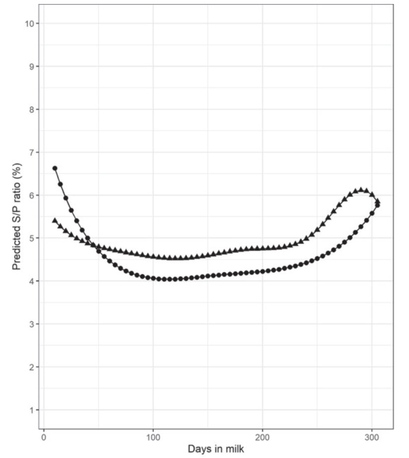
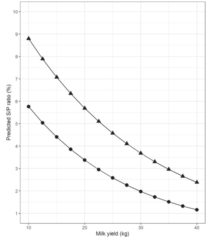
Comment: The above graphic from McAloon's paper illustrate that ELISAs for Johne’s disease done on milk samples are best done early or late in lactation. This study again shows a direct relationship between the magnitude of the ELISA value, called S/P value, and the yield of milk, i.e. higher ELISA S/P means lower milk yield (figure at the top of this page). Milk ELISAs are the least expensive testing option for dairy herds in most countries and are useful in controlling Johne’s disease and selecting cows for replacement.
MAP SURVIVAL IN CHEESE
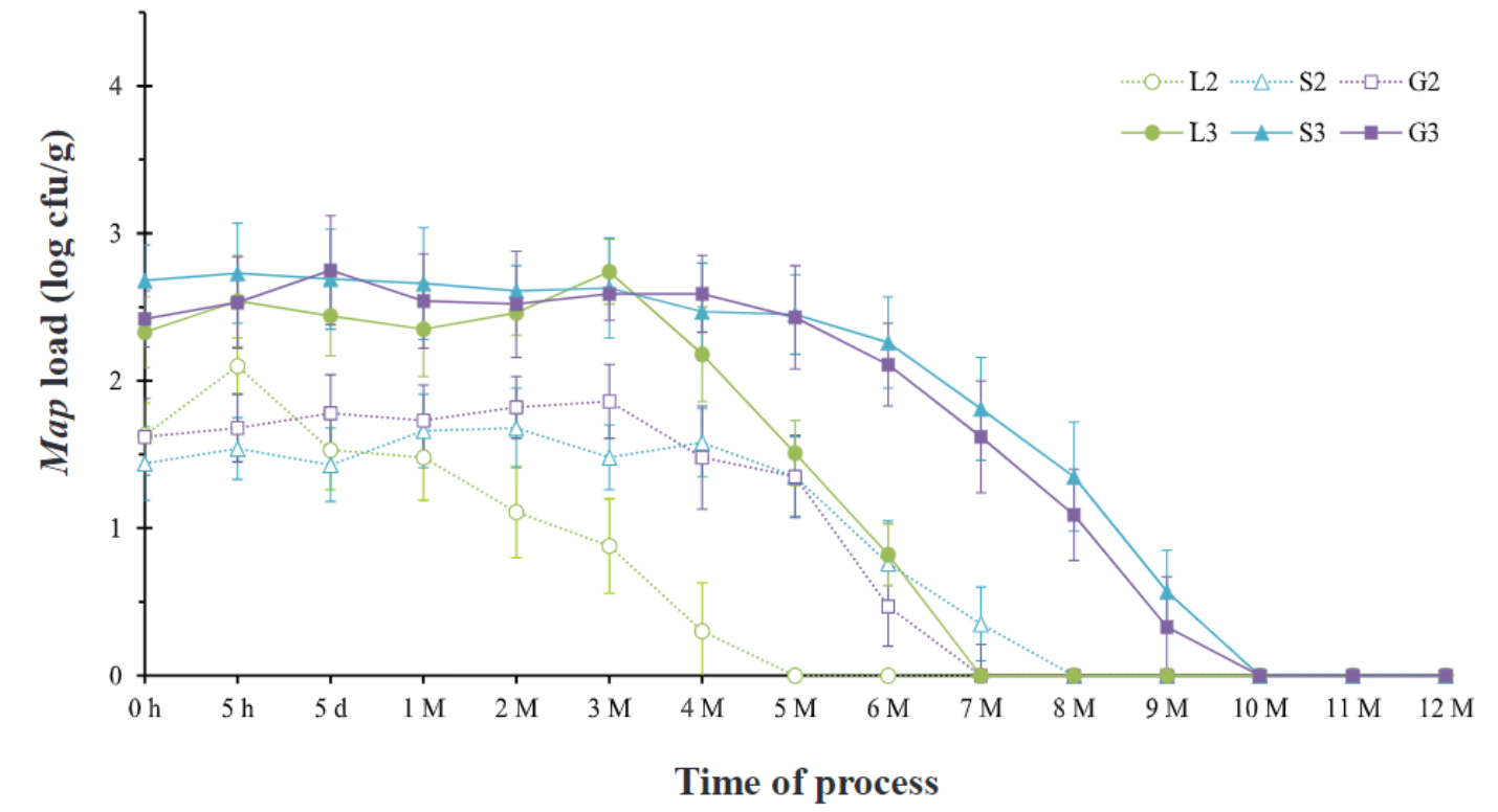
2020-08-07 14:48:07It takes months for MAP to die off during the manufacture of Lighvan cheese, a semi-hard raw milk cheese traditionally made from sheep's milk in Lighvan, a village in East Azerbaijan, Iran.

Shahram Hanifian from the Department of Food Science and Technology, Biotechnology Research Center, Tabriz Branch, Islamic Azad University, Tabriz, Iran has reported on the survival of MAP during the production and ripening of Lighvan cheese. His publication appears in the November 2020 issue of the journal LWT – Food Science and Technology. [Open Access]
Abstract
Mycobacterium avium subsp. paratuberculosis (Map) is an important animal pathogen with a worldwide distribution that may contribute to Crohn's disease in humans. This study aimed to investigate the behavior of Map in Lighvan cheese with special reference to the strains of Map, inoculum load, and storage time. One laboratory and two native strains of Map were inoculated (102 and 103 Map cell/mL) to cheese milk. The behavior of Map throughout the manufacture, ripening, and storage stage of Lighvan cheese tracked using propidium monoazide (PMA) qPCR and culture examination. According to the results, PMA-qPCR showed parallel outcomes in comparison with the culture assay. Besides, in all cheese batches Map was not affected during the ripening; however, throughout the storage period Map population decreased depending on the strain of Map. Trend analysis revealed no significant difference between the persistence of sheep and goat strains, but in all cheese batches, these native strains persisted significantly (p < 0.05) longer than the laboratory strain. In all treatments, the decreasing rate of Map found independent of the initial inoculum load. Nonetheless, in the cheese batches with higher initial inoculum, Map persisted two months longer.
Comment: Meta-analysis has shown that MAP is strongly associated with an inflammatory bowel disease called Crohn’s disease. Studies in Iran support this. Human exposure to MAP likely comes mostly from foods of animal-origin and cheese is one such product. This publication extends observations by others on cheese that MAP can survive extended times. Compared with Western countries, the incidence of IBD is lower in Iran; however, during the past decade, unofficial reports have claimed the incidence appears to be increasing (Aghazedh, 2005). MAP infections in Iranian animals, which are on the rise, may be party responsible.
Links of relevance:
IBD in Iran – a review.
Association of MAP with Crohn’s disease in Iran.
Paratuberculosis in Iranian goats.
Paratuberculosis in Iranian sheep.
Paratuberculosis in Iranian dairy cattle.
ECO-FRIENDLY WAY TO KILL MAP
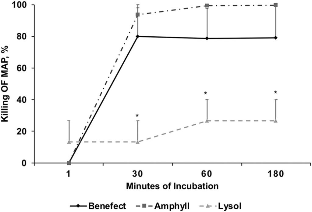
2020-08-02 15:49:18Dr. Judy Stabel and colleagues from the USDA-ARS-National Animal Disease Center, Ames, Iowa, USA compared new plant-based disinfectants to the more traditional phenolic and quaternary ammonium disinfectants in ability to kill MAP. Their publication appears in the September 2020 issue of the Journal of Microbiological Methods. This is a useful explanation of disinfectants for MAP and shows the contact time required for 100% killing, which is longer than for most microbes.

Abstract
Mycobacteria are difficult to kill due to the complexity of their cell wall. Further, Mycobacterium avium subsp. paratuberculosis (MAP) has one of the more elaborate cell wall compositions of all the mycobacteria. As a working pathogen within a research laboratory setting or as an environmental contaminant shed in the manure from infected animals, MAP is highly resistant to typical disinfectants. In the past, the most successful disinfectants to kill mycobacteria were based upon phenolics, harsh compounds that can break down the lipids within the cell wall. New disinfectants have been developed that are less toxic to the environment, however, it is unknown how well they perform compared to more traditional disinfectants. In the present study, we present comparative data on the utility of a commercial eco-friendly disinfectant, Benefect®, compared to Amphyl®, a phenolic-based disinfectant, and Lysol®, a quaternary ammonium-based disinfectant, to kill MAP in pure culture, tissues, and manure. Results demonstrated that Benefect was highly effective with up to 100% kill of MAP within 30 min in all experiments, paralleling results obtained with Amphyl. Lysol performed the most poorly, requiring longer contact times to kill MAP. These results suggest that natural, nontoxic ingredients can be used to disinfect even hearty pathogens such as MAP effectively, both within the laboratory and on-farm.
Comment: One of the more common questions from animal owners concerns disinfection of animal facilities after discovery of a MAP-infected animal. This new research gives a new and safer option for disinfection. Thank you Dr. Stabel!
ANTI-MAP FOR KIDS WITH CROHN’S
2020-07-25 15:48:07Drs. Agrawal, Hamblin, Clancy & Borody from the Research Department, Centre for Digestive Diseases, Five Dock, NSW, Australia describe successfully treating Crohn’s disease in children with anti-MAP antibiotics. Their publication, titled “Anti-Mycobacterial Antibiotic Therapy Induces Remission in Active Paediatric Crohn’s Disease” appears in the July 24, 2020 issue of Microorganisms.
Abstract
Crohn’s disease is increasing in incidence and prevalence in younger people and is of a particularly aggressive nature. One emerging treatment targets Mycobacterium avium paratuberculosis (MAP), an organism implicated in the causation of Crohn’s disease. This study reviewed a cohort of paediatric patients with active Crohn’s disease treated with Anti-Mycobacterial Antibiotic Therapy (AMAT). Sixteen paediatric patients, the majority of whom had failed conventional immunosuppressive therapy, were treated with AMAT. Endoscopic remission was scored using the Simple Endoscopic Score for Crohn’s Disease and clinical remission was assessed using the Weighted Paediatric Crohn’s Disease Activity Index (wPCDAI). Inflammatory blood markers were also routinely recorded. Patients were followed up clinically and endoscopically during treatment after an average of two months (range 1–6) and 17 months (range 2–49), respectively. A significant reduction in both scores assessing clinical improvement (p < 0.001) and mucosal healing (p < 0.0078) was observed at these timepoints; 47% of patients had achieved clinical remission and 63% endoscopic remission. Haemoglobin and serum inflammatory markers normalised for more than 50% of the cohort by six months of treatment. No adverse effects were reported throughout treatment. This is the first report of Anti-Mycobacterial Antibiotic Therapy offering a safe and efficacious therapy for paediatric patients with Crohn’s disease. Further larger randomised studies are required in order to validate these findings.
Author's Conclusions:
Despite a well-documented aggressive disease presentation and a recent exponential rise in the incidence of paediatric CD, fewer therapies are available in children when compared to the adult population. We examined the safety and efficacy of a combination antibiotic regime consisting of rifabutin, clofazimine and clarithromycin targeting MAP, a proposed contributing factor in the pathogenesis of CD. This study provides a valuable and novel proof of concept regarding the applicability of AMAT to a broad range of clinical presentations in paediatric Crohn’s disease patients. We report no significant side effects as a result of a dose escalated, sustained antibiotic regimen and suggest that AMAT may be more efficacious in the treatment of childhood onset compared with adult onset CD. Furthermore, we observed a notable trend, which proposes that the extent of prior immunosuppressive therapy may predict a longer treatment course with AMAT. Future targeted and more robust RCTs should also focus on sub-populations in terms of disease severity, extent of prior treatment and the time since initial diagnosis prior to the commencement of AMAT.
Comment: Read this for more about MAP in food and water and a list of 29 references to scientific literature. Given the weight of evidence and the severity and magnitude of potential human health problems, the precautionary principle suggests that it is time to take actions to limit “as low as reasonably achievable” human exposure to MAP. Only by controlling MAP in food producing animals can we stop the ongoing exposure of humans to MAP through the food supply.
FARMER & DVM VIEWS ON JD
2020-07-20 17:31:07Dr. Philip Robinson, School of Veterinary Medicine, College of Medical, Veterinary and Life Sciences, University of Glasgow used social science methods to explore farmer and veterinarian attitudes about Johne’s disease control in the UK. The publication titled “’They've got to be testing and doing something about it’: Farmer and veterinarian views on drivers for Johne’s disease control in dairy herds in England" appears in the September 2020 issue of Preventive Veterinary Medicine. The article also succinctly describes the UK’s National Johne’s Management Plan (NJMP).
This is one of the most interesting articles I have read is quite some time. It is filled with quotes from the dairy producers and veterinarians who were interviewed in conducting this study. There are many valuable lessons for how to motivate people and operate an effective national JD control program.
Must reading for everyone!
Abstract
There needs to be an understanding of the reasons why key stakeholders engage in disease control efforts if disease is to be successfully and sustainably controlled. It is increasingly recognised within veterinary epidemiology and policy making in animal health that these ‘people factors’ are important influences on the success or otherwise of animal disease control programmes. Research methodologies adopted from the social sciences offer ways to understand this important dimension through investigating the attitudes and opinions of the key actors involved. The study reported in this paper, based on qualitative interview research, investigates the views of dairy farmers and cattle veterinarians on the drivers and incentives for controlling Johne’s disease in English dairy herds. Twenty semi-structured interviews involving 17 dairy farmers and seven veterinarians were conducted in two dairy-intensive regions of England. The findings demonstrate the varied influences of veterinary advice and encouragement; appreciation of the economic cost of the disease at herd level; a voluntary national control plan; and fear of a future consumer food scare as the main reasons to engage in Johne’s disease control on dairy farms. The study demonstrates how a combination of a voluntary industry-led control scheme, compulsory participation through retailer and processor contractual requirements, and threats of reputational harm and market loss have strongly influenced farmer and veterinary behaviour in relation to Johne’s control without statutory involvement. The findings illustrate the importance of considering the political economy and societal impact of animal disease.
Author’s Conclusions
Johne’s disease control in dairy herds in England is not just about the trust that farmers have in the veterinary disease control advice of their veterinarians and whether it spurs action, or the relative economic merits of improving dairy herd health at the individual farm level. Rather, Johne’s control drivers would appear to be strongly influenced by wider industry concerns focused on the health implications of a possible causal association between MAP infection and Crohn’s disease in humans mediated through dairy cattle or dairy products. The findings illustrate the benefits of considering the wider political economy and potential societal impact of animal disease, and how these dimensions can also influence motivations for disease control, as argued in the introduction to this paper. The study raises interesting questions about the relationships between the roles and responsibilities for the control of endemic disease in food animals, food safety, and public and private goods in a global marketplace, and whether non-regulatory approaches by commercial private sector organisations are as effective, or even more effective, in raising animal health standards compared to statutory regulation. The relative socioeconomic and political merits of statutory, public-private partnerships, or privately funded animal health initiatives for endemic livestock diseases is an area which deserves further interdisciplinary research attention within the fields of veterinary epidemiology and animal health economics.
Commentary – some of my favorite quotes in the publication:
From and exasperated veterinarian:
‘I can think of a great example: this man has a [supermarket] contract, so he has to do Johne’s testing - it's obligatory for him. He loses clinical Johne’s cows hand over fist, and every single time I go there to look at a sick cow that's dropped its milk [I say] … “Is it on the Johne’s list?” [He says]:“I haven't looked.” So he does the recording because his milk buyer tells him he has to, but he's making no effort, even though he's losing … 5% of the herd a year to clinical Johne’s.’ (Int B04, veterinarian)
From a motivated veterinarian:
‘Yeah, it's just something I feel passionate about, and it's something I can see being a real issue. I also like my job, and I want my clients to be there in ten years’ time, and this could be a make or break for a lot of people.’ (Int B11, veterinarian)
From a farmer on his motivation:
‘AHDB (British farmer levy board) tell us it costs £1800 to rear a heifer to calving now. What’s the point in rearing a Johne’s heifer and spending £1800 on her when you might as well shoot it, or sell it and let someone else have the hassle? It doesn’t make sense - it’s hard enough farming as it is without knowingly rearing unhealthy animals.’ (Int A09, dairy farmer)
There are many many other interesting quotes and summary ideas about JD control in this important publication. Read it!
DAIRY: ELISA ON SERUM VS MILK
2020-07-13 18:30:15M.S.A. Faruk and colleagues from the Department of Animal Science and Technology, Sunchon National University in Korea describe a novel study on the kinetics of antibody production in dairy cattle. The article is titled: Longitudinal Study of Mycobacterium avium Subsp. paratuberculosis Antibody Kinetics in Dairy Cattle Using Sera and Milk throughout the Lactation Period and was published in the journal Veterinary Sciences 30 June 2020.
Abstract
Mycobacterium avium subsp. paratuberculosis (MAP) is the causative agent of Johne’s disease in dairy cattle populations around the world. The objective of this study was to evaluate MAP antibody kinetics in serum and milk samples throughout the lactation period in dairy cattle. The samples were collected simultaneously from eight MAP-positive and two healthy MAP-negative (control group) cows. The MAP antibody was detected by using serum and milk ELISA. The serum and milk MAP antibody titers fluctuated between the positive and negative cut-off values in this study. Specifically, cattle with low MAP antibody titer (<100) showed fluctuation between the cut-off values. Variable changes of MAP antibody titer were also observed after parturition. Between the serum and milk MAP antibody titers, there was a positive correlation (R2 = 0.5358) observed throughout the assessment period. The milk MAP ELISA test had low diagnostic performance in cows with low MAP titer due to its weak correlation (R2 = 0.0198). Finally, this study suggest that the periodic MAP ELISA test is recommended for the application of Johne’s eradication program due to the fluctuating nature of MAP antibody kinetics.
Conclusions of the authors
MAP antibody titers fluctuated in both serum and milk samples over the year, with the fluctuations occurring near the MAP-positive and MAP-negative cut-off borderline. These fluctuations make it difficult to diagnose a MAP-positive cow by only a single time measurement. The study result indicates that periodic MAP screening in a dairy herd is needed due to the fluctuating trend in MAP antibody level. In addition, the serum MAP antibody levels were gradually increased in high MAP antibody titer (>200) cows after parturition. However, some cows showed steady-state or decreasing trends in MAP antibody levels in low titer (<100) cows after parturition. There was a significant relationship between serum and milk sample results in cows with high MAP antibody titer (>200), but a weak relationship in cows with low (<100) MAP antibody titer. This weak agreement between serum and milk samples in low MAP antibody titer cows is indicative of low diagnostic performance of the milk MAP ELISA. Finally, the results of this study suggest that those farms applying Johne’s disease eradication programs should list cows as potentially MAP positive if their antibody titer lies near the Vet. Sci. 2020, 7, 81 9 of 10 cut-off value and periodic MAP ELISA testing is recommended due to the fluctuating nature of MAP antibody kinetics in dairy cattle.
Comment: Only 8 cows were included in the study and the authors state that they were “diagnosed as MAP-positive during the screening period”. It seems that the 8 cows were only selected based on ELISA results and did not have a paratuberculosis diagnosis confirmation by fecal PCR or culture. Without confirmation by MAP detection technology it remains unproven if all 8 cows were MAP-infected. As previously shown for serum ELISAs, the higher the ELISA S/P values the greater the confidence in the diagnosis (Collins-5_ELISA_Evaluation).
JOHNE'S SITUATION IN AFRICA
2020-07-06 17:57:04Research Review – OPEN ACCESS
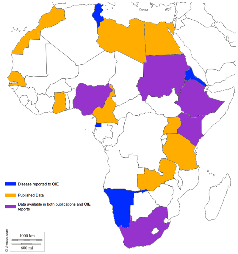
J.B. Okuni and 8 colleagues have published a review article describing the Johne’s disease situation in African countries and potential zoonotic concerns. Their article, titled Paratuberculosis: A Potential Zoonosis and a Neglected Disease in Africa appears in the July 5, 2020 issue of Microorganisms.
Abstract
The Mycobacterium avium subspecies paratuberculosis (MAP) is the causative agent of paratuberculosis, which is an economically important disease of ruminants. The zoonotic role of MAP in Crohn’s disease and, to a lesser extent, in ulcerative colitis, the two major forms of idiopathic inflammatory bowel disease (IIBD), has been debated for decades and evidence continues to mount in support of that hypothesis. The aim of this paper is to present a review of the current information on paratuberculosis in animals and the two major forms of IIBD in Africa. The occurrence, epidemiology, economic significance and “control of MAP and its involvement IIBD in Africa” are discussed. Although the occurrence of MAP is worldwide and has been documented in several African countries, the epidemiology and socioeconomic impacts remain undetermined and limited research information is available from the continent. At present, there are still significant knowledge gaps in all these areas as far as Africa is concerned. Due to the limited research on paratuberculosis in Africa, in spite of growing global concerns, it may rightfully be considered a neglected tropical disease with a potentially zoonotic role.

Conclusions
MAP poses a great challenge to the global livestock industry and is currently insidiously
spreading in Africa. Moreover, it could have possible impacts on human health across the continent. Given the fact that any known policies on this pathogen and most of the vital information required for instituting control and policy formulation are deficient, Africa remains a shadowy continent as far as this pathogen is concerned; therefore, paratuberculosis is, without any doubt, a neglected disease with possible zoonotic involvement in the African context. Given that the few studies undertaken on this disease have shown unfailing occurrence in several African countries, the disease is marching ahead of all stakeholders in the animal industry and needs to be closely studied. More attention in terms of funding and research needs to be given to Johne’s Disease in African countries.
ZOETIS ELISA KIT FOR CATTLE AND GOATS
2020-06-27 14:16:09 Zoetis produces an ELISA kit for Johne’s disease diagnosis that is USDA-licensed for use on cattle (bovine) and goat (caprine) samples. The kit is called SERELISA ParaTB Ab Mono Indirect. The publication providing accuracy analysis was published in SOJ Veterinary Sciences February 2016 [Open Access].
The last news posting incorrectly stated that VMRD was the only ELISA kit licensed for use on goat samples in the U.S. We apologize for this error. Outside the U.S. there are several other ELISA kits used on cattle and goats.
« Previous 1 … 7 8 9 10 11 … 19 Next »