
MAP IN PASTEURIZED MILK - IRAN
2021-01-06 01:00:51 Nasim Sadeghi from the Department of Food Hygiene, Faculty of Veterinary Medicine, Ferdowsi University of Mashhad, I.R. Iran and 2 colleagues have reported on the detection of MAP in pasteurized milk in northeastern Iran. Their research article appears in most recent issue of the Iranian Journal of Chemistry and Chemical Engineering.
Abstract
Mycobacterium avium subsp. paratuberculosis (MAP) is a gram-positive, small, acid-fast bacillus with high environmental resistance. In animals, especially ruminants, it leads to Paratuberculosis (PTB) or Johne's disease, which is chronic granulomatous enteritis. This bacterium as the main causative agent of Crohn's disease can be a serious threat to human health. This study aimed to detect MAP in pasteurized milk samples produced in Khorasan Razavi province, Iran, using Direct Nested PCR, PCR, and culture methods. In this study, 544 milk samples from Pasteurized Milk Production Companies were selected randomly during the 3-month period. DNA was extracted from milk fat after centrifugation. In order to identify the bacteria, Direct Nested PCR and PCR tests were applied using IS900 and f57, respectively. Furthermore, to detect viable MAP, positive samples resulted from Direct Nested PCR assays were cultured on Herrold's egg medium. For identification of mycobacterial isolates, all colonies were processed by PCR based on f57. A total of 544 pasteurized milk samples were assayed, and Mycobacterium paratuberculosis was detected in 39% of them by IS900 Nested PCR, and only 4.9% of samples were positive in the culture method. All the colonies were positive for the f57 using PCR. The results of this study indirectly indicated a high level of contamination of pasteurized milk to Mycobacterium paratuberculosis which is due to the large number of affected animals in livestock farms in Khorasan Razavi province. However, in comparison with the other researches, the low percentage of viable bacteria in pasteurized milk can be due to changes in temperature and time in pasteurizing systems of milk production companies in Khorasan Razavi province, Northeast of Iran.
Comment
When it comes to MAP, pasteurization is not perfect. The body of scientific evidence that viable (living) MAP occur in retail pasteurized dairy products continues to grow making this an important food safety issue. Rates of MAP detection by PCR methods, which do not distinguish living from dead MAP, are much higher than when using culture-based methods that detect only live MAP, as shown in the Iranian study. As new non-culture-based methods for detection of live MAP in dairy products are developed, I anticipate there will be even higher rates of viable MAP detection in dairy products. Also, the expanding paratuberculosis epidemic in animals globally results in steadily rising levels of MAP in all foods of animal-origin.
It is also important to consider that dead MAP in food may act as an allergen for some people potentially triggering inflammatory responses leading to diseases such as Type 1 Diabetes that are presently consider autoimmune disease. The cell walls of mycobacteria harbor some of the most potent immunogens known and have been used in Freund’s complete adjuvant to bolster immune responses to other antigens to produce high levels of antibodies in animals for decades.
Here is a partial list other published studies that have found live MAP in retail pasteurized milk:
- United Kingdom, Applied and Environmental Microbiology, May 2002.
- United States, Journal of Food Protection, May 2005.
- Czech Republic, Applied and Environmental Microbiology, March 2005.
- India, International Journal of Infectious Diseases, February 2010.
- Argentina, Brazilian Journal of Microbiology, July 2012.
- Brazil, Journal of Dairy Science, December 2012.
In closing…..
- Please forward the link to this news item to others you know who might be interested.
- Please subscribe to this website if you want to get regular emails about news items and content additions to the site.
- Please consider donating to help sustain this website.
MAP IN ZOO ANIMALS – REVIEW
2020-12-30 01:01:25Marco Roller and six colleagues in Germany and Brazil have published an excellent review article on MAP infections in zoo animals. This Open Access article appears in Frontiers in Veterinary Science (19 pages; 171 references).
ABSTRACT
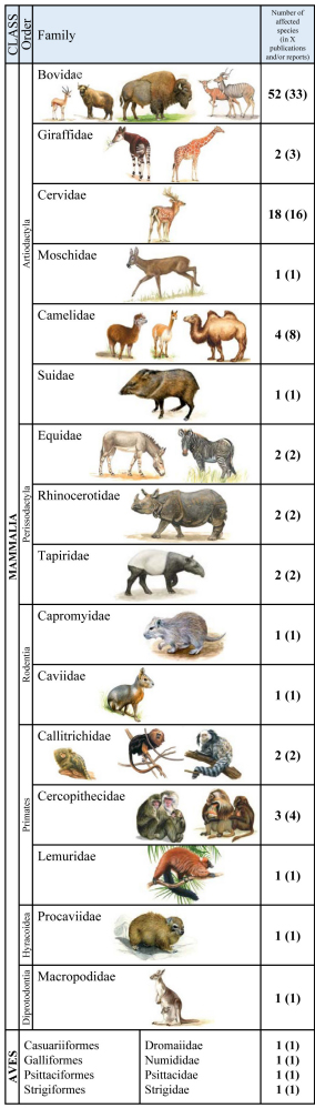 Mycobacterium avium subspecies paratuberculosis (MAP) is the causative agent of paratuberculosis (ParaTB or Johne’s disease), a contagious, chronic and typically fatal enteric disease of domestic and non-domestic ruminants. Clinically affected animals present wasting and emaciation. However, MAP can also infect non-ruminant animal species with less specific signs. Zoological gardens harbor various populations of diverse animal species, which are managed on limited space at higher than natural densities. Hence, they are predisposed to endemic trans-species pathogen distribution. Information about the incidence and prevalence of MAP infections in zoological gardens and the resulting potential threat to exotic and endangered species are rare. Due to unclear pathogenesis, chronicity of disease as well as the unknown cross-species accuracy of diagnostic tests, diagnosis and surveillance of MAP and ParaTB is challenging. Differentiation between uninfected shedders of ingested bacteria; subclinically infected individuals; and preclinically diseased animals, which may subsequently develop clinical signs after long incubation periods, is crucial for the interpretation of positive test results in animals and the resulting consequences in their management. This review summarizes published data from the current literature on occurrence of MAP infection and disease in susceptible and affected zoo animal species as well as the applied diagnostic methods and measures. Clinical signs indicative for ParaTB, pathological findings and reports on detection, transmission and epidemiology in zoo animals are included. Furthermore, case reports were re-evaluated for incorporation into accepted consistent terminologies and case definitions.
COMMENT
At least half of zoos in North America have had animals diagnosed with Johne’s disease. The infection spreads among institutions by the trade of animals; a practice that is essential for captive breeding programs. This led to a meeting of major zoo veterinarians and other zoo staff at the White Oak Conservation Center in Yulee, Florida in 1998. The 17-page proceedings of that meeting laid the groundwork for the control and prevention of Johne’s disease in zoological institutions. The White Oak proceedings are frequently cited in this publication by Rollo et al. Because these proceedings are hard to access, I have made it available here and on the website page about MAP infections of zoo ruminants.
MAP IN MANY HUMANS
2020-12-23 16:50:58Dr. J. Todd Kuenstner, Pathology and Laboratory Medicine, Lewis Katz School of Medicine, Philadelphia, PA, and colleagues from seven other institutions have reported on the detection of MAP in the blood of people with and without Crohn’s Disease by multiple laboratory assays. Their article appears in the most recent issue of Microorganisms [Open Access, 14 pages with 51 references].
![]()
Abstract
Mycobacterium avium subspecies paratuberculosis (MAP) has long been suspected to be involved in the etiology of Crohn’s disease (CD). An obligate intracellular pathogen, MAP persists and influences host macrophages. The primary goals of this study were to test new rapid culture methods for MAP in human subjects and to assess the degree of viable culturable MAP bacteremia in CD patients compared to controls. A secondary goal was to compare the efficacy of three culture methods plus a phage assay and four antibody assays performed in separate laboratories, to detect MAP from the parallel samples. Culture and serological MAP testing was performed blind on whole blood samples obtained from 201 subjects including 61 CD patients (two of the patients with CD had concurrent ulcerative colitis (UC)) and 140 non-CD controls (14 patients in this group had UC only).
Viable MAP bacteremia was detected in a significant number of study subjects across all groups. This included Pozzato culture (124/201 or 62% of all subjects, 35/61 or 57% of CD patients), Phage assay (113/201 or 56% of all subjects, 28/61 or 46% of CD patients), TiKa culture (64/201 or 32% of all subjects, 22/61 or 36% of CD patients) and MGIT culture (36/201 or 18% of all subjects, 15/61 or 25% of CD patients). A link between MAP detection and CD was observed with MGIT culture and one of the antibody methods (Hsp65) confirming previous studies. Other detection methods showed
no association between any of the groups tested. Nine subjects with a positive Phage assay (4/9) or MAP culture (5/9) were again positive with the Phage assay one year later. This study highlights viable MAP bacteremia is widespread in the study population including CD patients, those with other autoimmune conditions and asymptomatic healthy subjects.
Comment
While the high rate of MAP detection in human blood samples is shocking, it is not surprising. MAP has been proven capable of infecting a wide array of animal species, including nonhuman primates. The majority of MAP-infected animals, e.g., cattle, goats and sheep, are used for food. Since food safety regulatory agencies have not declared MAP to be a zoonotic pathogen, those MAP-infected animals, and their products such as milk, legally enter the food supply daily. Multiple studies have detected live MAP in retail pasteurized milk, cheese, and meat. Thus, humans are being exposed every day.
As the concluding sentence of this Dr. Kuenstner’s publication states: “as a minimum measure of best practice, the possibility that MAP is a zoonotic pathogen should prompt public health measures to better control JD and MAP spread into food and the environment by governments worldwide."
For more on MAP as a human pathogen visit the website of the Human Para Foundation, the organization who funded this study.
Read this for more evidence that MAP is a zoonotic pathogen.
Read this for more on MAP in food and water.
WHY I DO THIS
2020-12-18 01:06:40Once upon a time, about 17 years ago, there was a young Wisconsin girl named Lizi who was in 4H, a U.S. organization that provides experiences where young people learn by doing. Lizi decided to do a project to learn more about Johne's disease, something she had seen on her family's farm. She searched the web and found Johnes.org. Lizi used the information and images she found there to create a poster which she exhibited at the Lodi Agricultural Fair and Dane County Fair in Wisconsin, winning a blue ribbon for her efforts. Here is a picture of Lizi's poster.
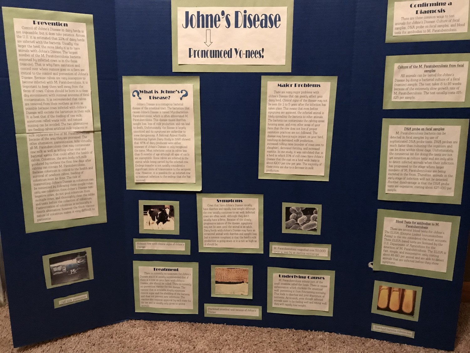
This story came to light when Lizi was in my Veterinary Bacteriology & Mycology class, Fall semester in 2017, learning (again) about Johne's disease. Lizi shared this picture of her poster and granted me permission to tell her story. 
The adjacent photo shows Lizi with a newborn calf on her family's farm. She graduated with her DVM degree from the University of Wisconsin School of Veterinary Medicine, May, 2020. She is pursuing a career in food animal medicine.
You never know what kind of impact knowledge sharing will have.
That is why I do this.
Michael T. Collins, DVM, PhD, DACVM
Comment: 4‑H is delivered by Cooperative Extension—a community of more than 100 public universities across the nation that provides experiences where young people learn by doing. For more than 100 years, 4‑H has welcomed young people of all beliefs and backgrounds, giving kids a voice to express who they are and how they make their lives and communities better.
If you feel you have seen this news posting before, you are not losing your mind. I originally posted this after Lizi was in my class and made me aware of our connection. In these tough times I feel that some heart-warming news like this is worth re-posting.
HAPPY BIRTHDAY DR. JOHNE!
2020-12-10 01:00:50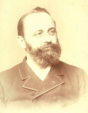
One-hundred eighty-one years ago, on December 10, 1839, Heinrich Albert Johne was born in Dresden, Germany. It seems fitting that Johnes.org celebrate, on this date, his lasting contribution to veterinary medicine.
Below you will find the story of his discovery, a brief biography, a little about the pathogen name, and the story of how I found this photo.
How it all started.
Dr. F. Harmes, a veterinarian in the Oldenburg region of Germany in 1895, had a client with a Guernsey cow that was doing poorly. Dr. Harmes’ preliminary diagnosis was intestinal tuberculosis (TB). TB in cattle was quite common in Germany then. But when he did the tuberculin skin test to confirm his diagnosis, the cow tested negative. So, the reason for the cow’s condition remained a mystery.
A few months later, the cow died. Curious as to what killed the cow, Dr. Harmes sent intestines and other tissues to the Pathology Unit at the veterinary school in Dresden. There the tissues were examined by Dr. Heinrich A. Johne, Professor of Pathology, and Dr. Langdon Frothingham, a visiting scientist from the Pathology Unit in Boston, Massachusetts.
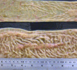 They observed that the small intestine was quite a bit thicker than expected and that lymph nodes near this thick intestine were enlarged. The photo at the right shows a normal intestine at the top and the intestine thickened due to Johne’s disease at the bottom. Lymphoid tissue, called Peyer’s Patches, are also quite prominent (the raised and slightly red tissue running long-ways down the center of the thickened intestine). Interestingly, Dalziel in 1913 saw the same kind of pathology when he removed a section of intestine from a person with Crohn’s disease remarking in his report that it resembled the cattle problem Dr. Johne had described.
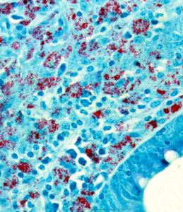 Using what at the time were newly developed histopathology techniques, parts of the intestine were “fixed” (pickled in formaldehyde), sliced into very thin sections, placed on a microscope slide, and stained with special dyes – known as an acid-fast stain - designed to help visualize bacteria of the type causing TB. Under the microscope, Drs. Johne and Frothingham saw that the intestinal wall was filled with inflammatory cells of the kind to be expected in TB (macrophages and lymphocytes – the blue-colored stuff in the photo). In addition, they saw abundant red-staining bacteria (which microbiologists call acid-fast bacteria) throughout the inflamed tissues. Basically, it looked just like intestinal TB. But, when a sample of the fresh infected tissue containing the red-staining bacteria was injected into guinea pigs, it didn’t cause TB. This took place shortly after Louis Pasteur had devised the “germ theory” of disease and before techniques for growing bacteria in the laboratory were widely available. Inoculating animals, therefore, was a routine way of detecting infectious microbes such as those that cause TB, and guinea pigs are quite susceptible to tuberculosis. So, the diagnosis on this cow remained a mystery.
Drs. Johne and Frothingham concluded that the disease seen in the very sick Guernsey cow was caused by a bacterium other than the one normally causing TB in cattle, namely Mycobacterium bovis. They speculated that perhaps the pathology was due to a related bacterial pathogen such as the one causing TB in birds, aptly named Mycobacterium avium. Considering their subject’s gross pathology, microscopic pathology (histopathology) and animal inoculation findings, they proposed the name "pseudotuberculous enteritis" for the disease; a designation meaning inflammation of the intestine resembling intestinal TB but not actually the same as intestinal TB – somehow different. Soon after publication of their report, veterinarians began reporting outbreaks of this curious intestinal malady among dairy cows in Denmark, The Netherlands and elsewhere in continental Europe.
More on Dr. Johne.
H.A. Johne was the son of a veterinarian. Twenty years later, he became a veterinarian himself and held a practice for the next seven years. From 1866-1876, he acted as district veterinary inspector. He was then appointed to a lectureship at the veterinary school in Dresden. For a teacher of veterinary medicine, he lectured in an unusually wide range of subjects: embryology, histology, obstetrics, exterior, physical diagnostics. In 1879, he was appointed professor of pathological anatomy and of general pathology. Later he also lectured on parasitology and methodical zoology, and he also started classes in such a new branch of research as bacteriology.
As a scientist, he concerned himself with tuberculosis, anthrax, rabies, glanders, actinomycosis, bothryomycosis among others. As a writer he left a wide literary production. His books were printed in a dozen editions. For many years he also edited “Zeitschrift tor Tiermedizin”, and acted as co-editor of “Rundschau auf dem Gebiet der Fleischbeschau”. In 1887, he visited Denmark, where he was nominated honorary member of the Danish Association of Veterinarians and decorated with the Order of Knight of the Dannebrog.
He was often guest of Professor B. Bang and his family, the flat of Bangs' is today the Veterinary History Museum, established in 1973 at the Royal Veterinary and Agricultural University in Copenhagen. His motto was: Duty Above All. With the distinguished array of titles: Geheim-Medizinalrat, Professor, Dr. med., Dr. med.vet.h.c. and Dr. phil., Heinrich Albert Johne retired in 1904, respected and honored by his many students and by foreign veterinary schools and societies. He died in 1910.
MAP
In 1912, in one of those curious discoveries by serendipity, Twort and Ingram discovered how to grow the cause of Johne’s disease in the laboratory and named this bacterial pathogen Mycobacterium enteritidis chronicae pseudotuberculosae bovis johne. Time and technology led to name changes and the cause of Johne’s disease is today known as Mycobacterium avium subspecies paratuberculosis or simply MAP. Johne’s disease, also called paratuberculosis, is now a disease of major global importance.
Dr. Johne photo credit
I found the photo of Dr. Johne was hanging in halls of the State Veterinary Serum Institute when I was on sabbatical working with Dr. J.B. Jørgensen at the State Veterinary Serum Laboratory, Copenhagen, Denmark. Together we were comparing new methods for culturing MAP from clinical samples. On my departure, Dr. Jørgensen gifted me a copy of this photo which hangs in my office and also appears on the Wikipedia page about Dr. Johne.
PS
For more historical events and people visit our history timeline.
POTENTIAL COST OF MAP IN MILK
2020-11-29 15:44:06L. Chiu and colleagues from Cornell University and the University of Illinois published a research article exploring the societal costs of MAP in the milk supply and a link of MAP to Crohn’s disease. Their work was published in the International Journal of Food System Dynamics [Open Access].
ABSTRACT
Welfare costs of a potential food shock were estimated by disseminating information to milk drinkers on the prevalence of Mycobacterium avium sub. paratuberculosis (MAP) in the U.S. milk supply, its potential linkage to Crohn’s disease in humans, and subsequent government intervention to minimize MAP in the milk supply. We found that 19.6% of milk consumers exposed to MAP information would stop milk consumption at current market prices, and that only 5% of those would return to their original milk consumption levels after the government intervention. Societal costs of the food shock after the intervention were estimated at $18.2 billion.
RELATED STUDY
A similar study by H. Groenendaal and Zagmutt was published in the Journal of Dairy Science in 2008. This article, not cited by Chiu, explored three scenarios developed based on the effectiveness of possible risk-mitigation strategies. As reported in their publication, “in the first scenario, it was assumed that an effective strategy exists; therefore, a negligible demand decrease in the consumption of dairy products was expected. In the second scenario, it was assumed that new risk mitigation would need to be implemented to minimize the health hazard for humans. In this case, a small milk demand decrease was expected, but larger demand decreases were also possible. The third scenario assumed that no fully effective risk mitigation was available, and this resulted in a considerable demand decrease and a potential reduction in milk supply as a result of regulatory measures. A milk demand reduction of 1 or 5% resulted in a reduction in consumer surplus of $600 million and $2.9 billion, and a reduction in dairy farm income of $270 million and $1.3 billion, respectively. A decrease in milk supply would cause a slight increase in total losses, but would cause the greatest losses to test-positive dairy farms. Given the current scientific knowledge about MAP and CD, we conclude that if a link were established, it is most likely that the first or second scenario would occur. Thus, consumer response and economic consequences to the discovery of such a link are expected to be limited, but could be large if the consumer's perception of risk is large or if risk-mitigation strategies were ineffective.”
COMMENT
Prevention pays! Dairy producers and processors should work to limit the potential impact of MAP on consumer acceptance of dairy products. The necessary diagnostic tools and knowledge on how to control MAP infections in dairy cattle are available. Some countries have implemented national control programs to help avert negative consumer reactions should medial science accept that MAP is a zoonotic pathogen. Actions to control MAP in dairy cattle can protect consumer confidence while also improving farm profitability and animal health and welfare.
JD CAUSES FALSE-POSITIVE TB TESTS IN CATTLE
2020-11-23 14:05:38 Mariana Assunção de Souza and 6 other Brazilian colleagues reported on the occurrence of Johne’s disease (paratuberculosis) in cattle that were necropsied based on a positive comparative cervical skin test for bovine tuberculosis (bTB). Their research article appears in the current issue of Acta Scientiae Veterinariae.
Abstract
Background: Bovine tuberculosis control programs are based on a standard diagnostic method, the intradermal test with purified protein derivatives, which is used to identify and eliminate diseased animals. Currently none of the tests available allow complete differentiation between infected and uninfected animals. The main limitations of the tests available are related to diagnostic sensitivity and specificity, which results in false-positive reactions due to the existence of cross infections, and also false-negative, inherent to the state of energy of some animals. The aim of this work was to study the intercurrence of paratuberculosis in tuberculosis reactive cattle by the comparative cervical test.
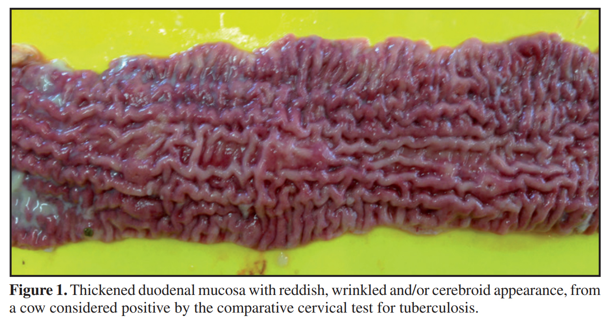
Materials, Methods & Results: Three hundred and thirty-four cattle were evaluated using the comparative cervical test (CCT) and serology for tuberculosis (TB) and paratuberculosis (PTB) ELISA IDEXX®. All of the animals testing positive by CCT were euthanized and necropsied. Fragments of lymph node, lung and intestine were collected and analyzed using histopathological techniques, with staining by Hematoxylin-Eosin (HE). Samples of lung and lymph nodes (retropharyngeal, submandibular, cervical and mediastinal) of the animals testing positive by CCT were evaluated using qPRC for M. bovis, and intestinal and mesenteric lymph nodes using PCR for PTB. Of the 334 cattle evaluated using the comparative cervical test, 16 were considered positive. No lesions suggestive of tuberculosis were found in the macroscopic inspection of the carcasses. The most evident anatomical and pathological finding was a thickening of intestinal mucosa, found in 12 of the 16 cattle submitted to necropsy. No microscopic lesions suggestive of TB were identified nor was the presence of M. bovis detected by qPCR. The main histopathological findings were observed in the small intestine and mesenteric lymph nodes and identified as enteritis, lymphangitis, lymphangiectasia and granulomatous lymphadenitis. In the intestine the changes are characterized by dilated and inflamed lymphatic vessels and intense inflammatory infiltrate on the mucosa and submucosa. Of the 334 serum samples evaluated, the M. bovis ELISA Antibody Test (IDEXX®) identified 17 positive animals. All the cattle considered positive by M. bovis ELISA were considered negative by CCT. In the samples from nine animals (9/16), DNA from M. avium subsp. paratuberculosis (MAP) was identified and in twelve carcasses (12/16) lesions characteristic of PTB were found, which were subsequently confirmed by histopathological techniques. In another nine animals of the herd anti-MAP antibodies were detected. None of those that tested positive by PTB ELISA were reactive by CCT.
Discussion: Animals considered positive by TB ELISA that were not positive in the intradermal test does not mischaracterize the clinical picture of the disease. Considering the inverse relationship between cell-mediated and humoral responses to M. bovis, the intradermal test and the serological tests are designed to measure different immunological responses, which develop during different stages of infection. The progress of the cellular immunological response to humoral immunity occurs in the most advanced stages of tuberculosis. Of the 16 cattle considered positive by CCT, 12 animals presented macroscopic and histological lesions suggestive of PTB and DNA from MAP was detected in nine. Although it is the official test for the control of TB in different countries, the intradermal test with PPD has presented limitations, primarily related to specificity. M. avium subsp. Paratuberculosis is considered the main cause of false positive reactions in the intradermal test. The PPD bacterial extract is a complex mixture of proteins, lipids, sugars and nucleic acids, and many of these components are also shared by numerous species of mycobacteria (tuberculous or not).

The Brazilian study illustrates the interaction of MAP and M. bovis, the cause of bovine tuberculosis (bTB), as a confounder when trying to use standard immune-diagnostic tests for bTB. Other authors have found similar results. Out October 30 news posting on this site discussed the need to wait at least 60 days after skin testing cattle for bTB before using serological tests (ELISA) for Johne’s disease (JD) in order to avoid false-positive JD blood tests.
In a different but related context, other authors have likewise noted this interaction between mycobacterial diseases such as TB, leprosy and those caused by non-tuberculosis mycobacteria (NTM). The most common NTM infections are due to bacteria in the Mycobacterium Avium Complex (MAC) which includes the cause of JD, i.e. MAP.
In a letter published in the journal Inflammatory Bowel Diseases in 2008, Dr. Marcel Behr from McGill University Health Centre, Montreal, Canada highlighted instances where one mycobacterial infection interferes with another. One example he cited is that TB and leprosy (caused by Mycobacterium leprae) behaved as antagonistic epidemics – one infection blocks the other. When human TB is controlled, as it has in most developed countries, and when BCG vaccination of humans stops, other mycobacterial infections rise in prevalence. The theory is that the absence of one mycobacterial infection, e.g. TB, causes an “immunological void”, i.e. an increased susceptibility of humans to other mycobacteria that now find more susceptible, immunologically naïve, hosts. Note: BCG is a live non-virulent vaccine used to control TB in humans in countries where the disease is endemic. This vaccine was originally derived from M. bovis.
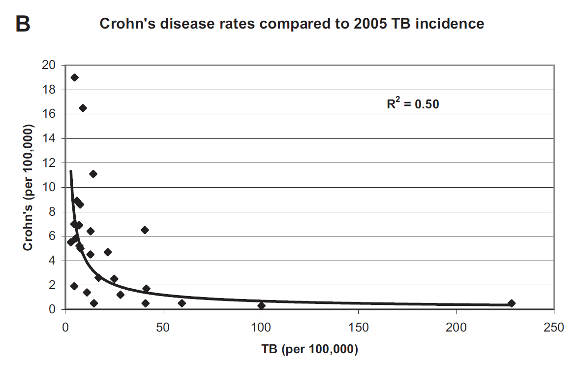
Because MAP is strongly associated with Crohn’s disease, Dr. Behr noted that Crohn’s disease (CD) was more common in countries where TB was less common. Below is the graphic he published illustrating inverse association between TB incidence and CD incidence. This is yet another piece of evidence for mycobacterial, e.g. MAP, involvement in CD.
Related to this is the review article by Dr. Dow (15 pages with 173 references) published in February 2020 on the influence of BCG vaccination on a variety of autoimmune diseases. In that article he states: "MAP has been associated with an increasingly long list of inflammatory/autoimmune diseases: Crohn's disease, sarcoidosis, Blau syndrome, Hashimoto’s thyroiditis, autoimmune diabetes (T1D), multiple sclerosis (MS), rheumatoid arthritis, lupus and Parkinson’s disease. Epidemiologic evidence points to BCG providing a “heterologous” protective effect on assorted autoimmune diseases; studies using BCG vaccination for T1D and MS have shown benefit in these diseases." His article proposes that the positive response to BCG in T1D and MS is due to a mitigating action of BCG upon MAP infections.
For more on the potential of MAP to cause Crohn’s disease visit this page of our site. Or listen to a 10-minute presentation titled: MAP is a zoonotic pathogen.
15-ICP POSTPONED AGAIN
2020-11-20 16:09:49
In March 2020, in response to the global pandemic brought about by the COVID-19 virus the Committee of the 15th International Association for Paratuberculosis Colloquium 2020 made the hard decision to postpone ICP 2020 until 2021. The event was originally scheduled for 14th -18th June, 2020 in Dublin Castle. The 15th International Colloquium for Paratuberculosis was then re-scheduled for the 6th – 9th April 2021.
Last month the Local Organizing Committee (LOC) was faced with the decision of having to postpone again or to have a virtual conference next April. It was decided by the LOC to go for a live conference in June 2022. The committee was delighted that the organizers of the 16th ICP in Jaipur, India agreed to this and have postponed their Colloquium until 2024.
The ICP Committee invites you to the 15th IAP Colloquium in Dublin, Ireland in June 2022. Delegates attending the conference can be assured of a productive and memorable colloquium, discover Irish heritage, culture and music and of course, experience the world-renowned hospitality of Ireland.
OJD REVISITED – AUSTRALIA
2020-11-13 17:54:34
Dr. Peter Windsor and Dr. Richard Whittington published a review article on ovine Johne’s disease (OJD) control in Australia. This excellent review article appears in the journal Animals and is Open Access. It nicely reviews the pathogenesis and control of paratuberculosis in sheep, but the most interesting part of the article addresses the top 10 subjects of misinformation about OJD in Australia.
Abstract
OJD is no longer the serious animal health issue that it was for many Australian rural communities a decade and a half ago. Despite declining OJD prevalence as determined by abattoir surveillance, the disease continues to spread, with OJD extension programs required to continually address the misinformation promulgated by some disaffected producers as new areas have become affected. Improved regional and on-farm biosecurity, including the introduction of a risk-based trading system, may have contributed to improved attitudes to OJD control, although attitudinal differences between OJD endemic areas and where the disease is not well established remain. Declines in on-farm OJD prevalence are almost certainly attributable to the widespread uptake of vaccination programs, although encouraging the ongoing use of vaccination to prevent recrudescence and improved biosecurity when mortalities disappear, remains challenging. Vaccination has provided a robust strategy for managing OJD and contributed significantly to the health of Australian sheep and the lives of producers with affected properties. As vaccination offers a pathway to reduce the risk of MAP infection entering the human food chain from small ruminant products, it should be more widely adopted globally, accompanied by research efforts to improve efficacy and importantly, the safety of vaccination to both operators and livestock.
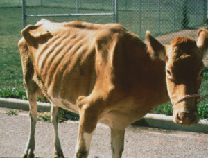
Comment: Not all countries, such as the U.S., have access to the vaccine for paratuberculosis that is used in Australia. For pictures of sheep before and after developing clinical Johne’s disease can be found here.
JD IN SCIMITAR ORYX
2020-11-06 17:13:38Claudio Pigoli and colleagues report on multiple cases of Johne’s disease in captive scimitar-horned oryx at a zoo in Italy. Their research article was published in the journal Animals and is Open Access. Genomic analysis shows that the MAP strains recovered from the Oryx are closely related to those isolated from cattle in Italy: evidence of ongoing spillover from domestic livestock to wildlife and animals in zoos.

Abstract
Paratuberculosis, a chronic disease caused by Mycobacterium avium subsp. paratuberculosis (MAP), in ten scimitar-horned oryxes (SHOs) hosted in an Italian zoological park and originating from a Slovakian flock, was documented by pathology, molecular, cultural, and serological testing. The infection origin in this threatened species was also investigated by genomic analyses. Following the death of six of the 10 SHOs, serial investigations of dead and alive animals were performed. Necropsy, carried out on five out of six animals, identified intestinal thickening and mesenteric lymphadenomegaly in one of the animals. Histopathology (5/6) revealed lepromatous (2/5) and tuberculoid (2/5) intestinal forms or lack of lesions (1/5). Ziehl-Neelsen and immunohistochemistry stains identified two multibacillary, two paucibacillary forms, and one negative case. MAP was identified by quantitative PCR (qPCR) in tissue samples in five out of five SHOs and was microbiologically isolated from two of the three animals whose fresh tissue samples were available. Fecal samples were collected in four of the six dead animals: all four resulted positive to qPCR and in MAP was isolated in three. ELISA identified MAP-specific antibodies in three of the five dead animals whose serum was available. qPCR identified MAP in the freshly deposited feces of two out of the four alive animals. From the feces of these two animals, MAP was microbiologically isolated in one case. All isolates were classified as MAP type C and profiled as INMV2 and MVS27 by molecular analysis. Genomic analysis of a field isolate revealed clustering with a European clade but was more similar to Italian than East European isolates. Our findings underline that paratuberculosis should always be considered in zoological parks in which endangered species are hosted. Infection can be subclinical, and multiple combined testing techniques may be necessary.
Author’s Conclusions
Our results underline the importance of considering paratuberculosis in zoological parks, where endangered species are often hosted. Paratuberculosis could represent a risk for the conservation of rare animals, and it is essential to include it in the panel of diagnostic tests to be performed on hosted animals. We also suggest testing dead animals, in which different diagnostic approaches are combined, with the final aim of fully elucidating the causes of death and defining their health status regarding paratuberculosis. WGA can help to trace the origin of infections, particularly in the case of moved animals. This study reports the first genome of an MAP strain isolated from SHOs and shows that the strain likely derived from the Italian cattle livestock, in which MAP is endemic.
For more on Johne’s disease in zoo animals check out this page of our website.
« Previous 1 … 5 6 7 8 9 … 19 Next »